


Peptide Absorption: 8 Factors That Shape Research Use
Research use demands understanding 8 factors that shape peptide absorption: pH, proteases, mucus, size, hydrophilicity, structure, transporters, and formulation.
📋 Table of Contents
For research use, peptide absorption is governed by eight interacting variables: GI pH, proteolytic degradation, intestinal mucus, molecular size, hydrophilicity and charge, primary and higher-order structure, transporter uptake such as PepT1, and formulation strategy. Survival across the gastrointestinal tract plus transport across the intestinal epithelium determines whether a peptide reaches tissue or circulation; no single factor predicts absorption alone.
Key takeaways
- Oral peptide absorption for research use is a multi-barrier problem, not a single permeability issue: GI degradation, poor epithelial permeability, peptide structure, and first-pass hepatic metabolism combine.
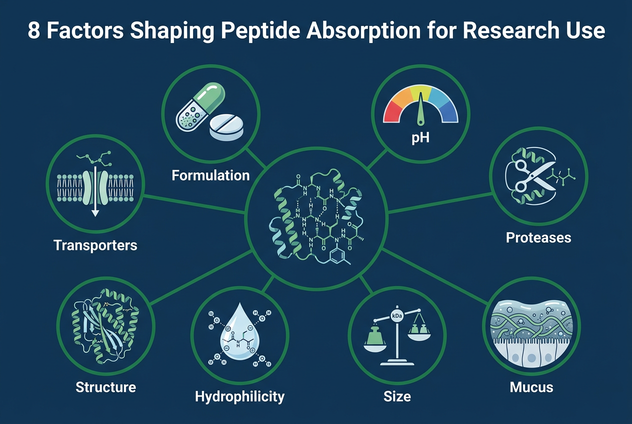
- The eight decisive factors are GI pH, proteases, mucus, molecular size, hydrophilicity, structure, transporter uptake (PepT1), and formulation strategy.
- Evaluation sequence matters for research use: test luminal stability first, epithelial transport second, systemic exposure third.
- Common oral-delivery tactics include pH modulation, enzyme inhibitors, permeability enhancers, and structure-focused design changes, each with trade-offs.
- A peptide can be active in vitro yet fail in vivo if unstable, too hydrophilic, or unable to cross the gut barrier; absorption is survival plus transport, not dose swallowed.
What does peptide absorption actually mean in research use?
In research use, peptide absorption means a peptide crosses a biological barrier—usually the intestinal epithelium—and reaches tissue or circulation. Peptides Library and recent PMC reviews treat this as survival plus transport, not simply the dose swallowed.
In research use, absorption is the movement step only. Bioavailability is broader: it includes absorption, presystemic loss, and the fraction of unchanged peptide that reaches systemic circulation. That distinction matters because a peptide detected in the gut lumen has been administered, not absorbed.
A common misconception is that oral exposure begins once a peptide enters the small intestine. In practice, the peptide must remain intact long enough to approach the epithelial surface, move through mucus, interact with transport pathways or membranes, and then survive early metabolism. If any one step fails, measured systemic levels can remain negligible.
Why is oral peptide absorption so difficult in research use?
Oral peptide absorption is difficult in research use because proteases, acidic gastric pH, mucus, and tight epithelial membranes act together. The small intestine and liver create a sequence of barriers rather than one isolated bottleneck.
Recent PMC reviews and FDA discussion of peptide drug development point to the same core problem (PubMed search on oral peptide absorption barriers, NIH NLM peptide drug delivery resources): peptides are often large, polar, and enzymatically fragile. That means they face degradation in the lumen, poor transcellular diffusion across membranes, and limited paracellular passage because tight junctions restrict movement of larger hydrophilic molecules. A frequent research error is treating oral absorption as a membrane problem alone.
Peptides Library emphasizes absorption-related characteristics rather than brand endorsements, a useful reminder that oral peptide performance starts with barrier biology.
Timing also matters. If a peptide is degraded in the stomach or proximal small intestine, transporter access later in the tract may never occur. If it survives but enters portal blood only briefly, first-pass hepatic metabolism can cut systemic exposure even more. So the limiting factor is often whichever barrier acts first and most strongly.
💡 Key Takeaway: Why is oral peptide absorption so difficult in research use?
What are the 8 factors that shape peptide absorption for research use?
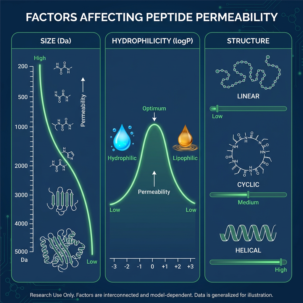
Eight variables matter most for research use: pH, proteases, mucus, peptide size, hydrophilicity, structure, transporter uptake including PepT1, and formulation strategy.
These factors interact rather than operate independently. A favorable change in one area can help or hurt another. Increasing lipophilicity may improve membrane interaction, for example, but it can also change solubility, aggregation behavior, or clearance.
| Factor | Effect for research use | Key detail |
|—|—|—|
| GI pH | Alters ionization, folding, and chemical stability | Stomach acid to near-neutral intestinal conditions |
| Proteolytic degradation | Rapid cleavage of susceptible sequences | Pepsin, pancreatic enzymes, brush-border peptidases |
| Intestinal mucus | Slows diffusion, blocks access to epithelial transport sites | Selective barrier for larger, charged, aggregation-prone peptides |
| Molecular size | Larger peptides cross membranes less readily | Collagen hydrolysates often 2,000–5,000 Daltons |
| Hydrophilicity and charge | Highly polar peptides have limited passive permeability | Multiple charges at intestinal pH reduce diffusion |
| Primary and higher-order structure | Sequence, conformation, protease-sensitive sites influence stability and transport | Cyclic motifs and resistant sequences help stability but not always transport |
| Transporter uptake | PepT1 may assist uptake of certain small bioactive peptides | Brush-border transporter in small intestine |
| Formulation strategy | pH modulation, enzyme inhibitors, permeability enhancers, protective dosage forms | Improves exposure but each has trade-offs |
The practical takeaway for research use is simple: no single factor predicts absorption well by itself. Researchers get better answers when they model the peptide, the barrier, and the dosage strategy together.
How does GI pH change peptide stability step by step for research use?
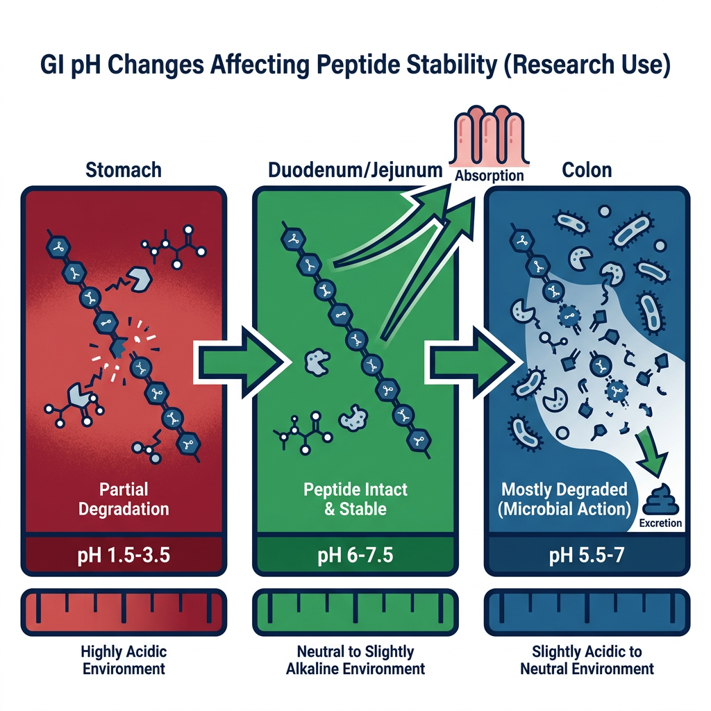
For research use, GI pH alters ionization and bond stability in stages, from stomach acid to near-neutral small intestine conditions.
First, the stomach exposes the peptide to a low-pH environment. That can change charge distribution, alter folding, and make some sequences more vulnerable to acid-assisted degradation or enzyme attack. Not every peptide is destroyed there, but many are destabilized enough that downstream survival drops.
Next, the peptide enters the small intestine, where pH rises and the enzyme environment changes. A higher pH may reduce some acid-related stress, yet it also coincides with pancreatic proteases that can hydrolyze peptide bonds efficiently. This is why pH alone never tells the full story.
Last, formulation strategies try to control when and where the peptide is released. Enteric protection or pH modulation may delay exposure to gastric acid, but that does not remove intestinal enzymatic pressure. A common misconception in research use is that buffering the stomach solves oral peptide delivery. It only addresses one barrier.
How do enzymes and the mucus layer reduce peptide uptake step by step for research use?
In research use, proteases and mucus reduce uptake sequentially: luminal enzymes cut peptides first, then mucus slows diffusion toward the brush border.
The sequence starts with enzymatic exposure in the lumen. Pepsin in the stomach, followed by intestinal and brush-border proteases, can reduce intact peptide concentration before it ever reaches the epithelial surface. That is why two peptides with similar receptor activity can show very different in vivo behavior.
Peptides Library notes that collagen hydrolysates are often 2,000 to 5,000 Daltons, a concrete example of how peptide size can change intestinal absorption behavior in research use.
After that, the mucus layer becomes important. Mucus is not just a passive coating. It acts as a selective diffusion barrier, especially for larger, charged, or aggregation-prone molecules. Pro tip for research use: do not treat mucus penetration and protease resistance as the same property. A peptide can resist cleavage reasonably well and still fail to reach the brush border in useful amounts.
💡 Key Takeaway: How do enzymes and the mucus layer reduce peptide uptake step by step for research use?
PepT1 vs passive diffusion: which absorption route matters more for research use?
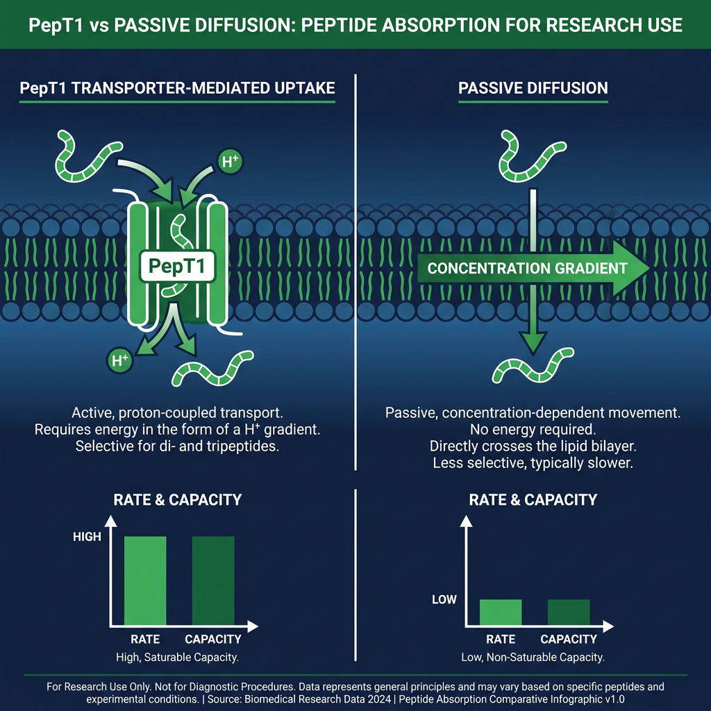
For research use, PepT1 often matters more than passive diffusion for small peptide uptake, while larger hydrophilic peptides rarely cross membranes efficiently on their own.
PepT1 is a brush-border transporter in the small intestine associated with uptake of dipeptides, tripeptides, and some bioactive peptide-like compounds. In research use, transporter access can be a major advantage because passive transcellular diffusion is usually poor for polar peptides.
Passive diffusion, by contrast, favors smaller and less polar molecules with sufficient membrane affinity. Many peptides do not meet those criteria. If a peptide is highly hydrophilic and carries multiple charges at intestinal pH, diffusion through the lipid bilayer is limited.
A useful check for research use is this: if the peptide is too large for likely transporter recognition and too polar for passive diffusion, then oral absorption will probably depend heavily on formulation tricks or remain intrinsically low. A common misconception is that all small peptides can use PepT1 efficiently. Transporter recognition is selective, not automatic.
For related transport pathways, the Cell Penetrating Peptides for Research | Peptides Library reference discusses mechanisms that complement oral absorption studies in research use.
How do peptide size, hydrophilicity, and structure affect permeability in research use?
In research use, large size, high hydrophilicity, and flexible structure usually lower membrane permeability; cyclic motifs and protease-resistant sequences often improve stability but not always transport.
Molecular size is a first filter. Larger peptides generally cross membranes less readily than smaller ones, partly because passive transcellular diffusion requires some membrane interaction that bulky polar molecules lack. The collagen hydrolysate example—2,000 to 5,000 Daltons—shows how size alone can shift intestinal absorption behavior.
Hydrophilicity and charge matter next. Highly polar peptides have limited passive membrane permeability. Multiple charges at intestinal pH make the molecule less likely to partition into the lipid bilayer, so paracellular transport becomes the only remaining route, but tight junctions restrict that path for larger molecules.
Structure influences both stability and transport. Primary sequence, conformation, and protease-sensitive sites determine how quickly a peptide is cleaved and whether it can adopt a transport-competent shape. Cyclic motifs and resistant sequences often improve survival, but they do not guarantee membrane crossing. A stable peptide can still be too polar or too large to reach systemic circulation in research use.
For laboratory reference material, the Peptide Library: Research-Grade Peptides for Laboratories page provides context on how structural variation affects absorption-related behavior in research use.
What formulation strategies improve peptide absorption in research use?
For research use, formulation strategies aim to improve exposure by addressing one or more barriers: pH modulation, enzyme inhibitors, permeability enhancers, and protective dosage forms.
pH modulation and enteric protection delay exposure to gastric acid. That can preserve acid-labile peptides, but it does not remove intestinal enzymatic pressure. A peptide that survives the stomach may still be cleaved by pancreatic or brush-border proteases in the small intestine.
Enzyme inhibitors reduce proteolytic degradation, but selectivity and reproducibility are common concerns in research use. Broad inhibition can alter normal digestive processes in animal models and complicate interpretation of absorption data.
Permeability enhancers act on tight junctions or membrane fluidity. They may increase paracellular or transcellular transport, but they can also compromise barrier integrity, leading to variable systemic exposure and potential off-target effects.
Protective dosage forms—such as nanoparticles, liposomes, or hydrogel matrices—can shield peptides from enzymes and mucus while controlling release. The trade-off is formulation complexity, batch variability, and the need to confirm that the dosage form itself does not alter the peptide’s research use stability profile.
For a broader reference set, the Peptide Research Library: A Complete Reference Guide covers absorption-related characteristics and formulation variables across peptide classes for research use.
💡 Key Takeaway: What formulation strategies improve peptide absorption in research use?
How should researchers evaluate peptide absorption in sequence for research use?
For research use, evaluate absorption in sequence: luminal stability first, epithelial transport second, systemic exposure third. That order matters because many peptide discussions mix up absorption, bioavailability, and efficacy.
Luminal stability testing should measure intact peptide remaining after exposure to simulated gastric fluid, simulated intestinal fluid, or relevant protease cocktails. If degradation is rapid, later transport measurements will not reflect the peptide’s true potential.
Epithelial transport should follow only after stability is characterized. Use Caco-2 monolayers, Ussing chambers, or ex vivo intestinal segments to measure apical-to-basolateral flux. A peptide that survives the lumen but cannot cross the epithelium will still show negligible systemic exposure.
Systemic exposure comes last. In animal models, measure portal and systemic concentrations after oral administration. First-pass hepatic metabolism can cut systemic exposure even when intestinal absorption occurs, so portal sampling helps separate absorption from presystemic loss.
Checklist for research use:
- [ ] Measure luminal stability before transport assays.
- [ ] Confirm intact peptide concentration after protease exposure.
- [ ] Test epithelial permeability with a validated barrier model.
- [ ] Measure portal and systemic exposure in the same study.
- [ ] Report absorption, bioavailability, and efficacy as separate endpoints.
For laboratory reference material, the Peptide Library: Research-Grade Peptides for Laboratories page provides context on how structural variation affects absorption-related behavior in research use.
What are common misconceptions about peptide absorption in research use?
Several misconceptions distort peptide absorption studies in research use. The first is that oral exposure begins once a peptide enters the small intestine. In practice, the peptide must survive degradation, cross mucus, and interact with transport pathways before absorption can occur.
A second misconception is that all small peptides can use PepT1 efficiently. Transporter recognition is selective, not automatic. Dipeptides and tripeptides with specific structural features are favored, but many small peptides are not substrates.
A third misconception is that buffering the stomach solves oral peptide delivery. Gastric acid is only one barrier. Intestinal proteases, mucus, tight junctions, and first-pass metabolism remain intact after pH modulation.
A fourth misconception is that absorption equals bioavailability. Absorption is the movement step; bioavailability includes presystemic loss and the fraction of unchanged peptide reaching systemic circulation. A peptide can be absorbed and still show low bioavailability due to hepatic metabolism.
A fifth misconception is that in vitro activity predicts in vivo absorption. A peptide can be biologically active in vitro and still perform poorly in vivo if it is unstable, too hydrophilic, or unable to cross the gut barrier efficiently in research use.
How does first-pass hepatic metabolism affect peptide absorption?
First-pass hepatic metabolism cuts systemic exposure even when intestinal absorption occurs. Portal blood concentrations can be high while systemic levels remain low, so measuring only systemic exposure underestimates absorption.
After a peptide crosses the intestinal epithelium, it enters portal blood and travels to the liver before reaching general circulation. Hepatic enzymes can metabolize a fraction of the absorbed peptide, reducing the amount that appears in peripheral blood. This presystemic loss is part of bioavailability but not part of absorption itself.
The distinction matters. If a study measures only systemic concentrations after oral dosing, it cannot tell whether low exposure resulted from poor intestinal uptake or rapid hepatic clearance. Portal vein sampling in animal models helps separate these two barriers.
A common error is to attribute low systemic peptide levels solely to poor oral absorption. First-pass metabolism may be the dominant reason in some cases, especially for peptides that are substrates for hepatic peptidases. Evaluating absorption in sequence—luminal stability, epithelial transport, systemic exposure—prevents this conflation.
💡 Key Takeaway: How does first-pass hepatic metabolism affect peptide absorption?
What role does peptide structure play in protease resistance?
Peptide structure determines both stability and transport. Primary sequence, higher-order conformation, and protease-sensitive sites directly influence how quickly luminal and brush-border enzymes can cleave a peptide.
Susceptible sequences—such as those containing certain basic or hydrophobic residues—may be cleaved rapidly by trypsin, chymotrypsin, or brush-border peptidases. Cyclization, N-methylation, or D-amino acid substitution can reduce protease recognition, but these modifications may also alter receptor binding or transporter recognition.
Conformation matters too. A flexible linear peptide exposes more cleavage sites than a constrained cyclic or stapled peptide. However, increased rigidity can reduce paracellular passage if the molecule becomes too large or too rigid to diffuse through tight junction-associated spaces.
Stability and transport are not interchangeable. A protease-resistant peptide may still fail to cross the epithelium if it remains highly hydrophilic or too large for passive diffusion. Researchers should evaluate both stability and permeability before concluding that structural modification improved absorption.
Which models best capture absorption barriers?
No single model captures all absorption barriers, so a tiered approach is more informative. Simulated gastric and intestinal fluids assess luminal stability; Caco-2 monolayers and Ussing chambers assess epithelial transport; animal models assess systemic exposure and first-pass metabolism.
Caco-2 monolayers are widely used for permeability screening, but they overexpress some transporters and lack a full mucus layer, so results may not match in vivo behavior. Ussing chambers preserve intestinal tissue architecture, including mucus and some enzyme activity, but viability is limited to a few hours.
Ex vivo intestinal segments retain mucus, tight junctions, and regional transporter expression better than cell lines, yet they still lack blood flow and hepatic clearance. In vivo rodent or porcine models provide the most complete picture, but species differences in peptide transporters and metabolism can limit translation to other laboratory contexts.
A practical sequence is to combine models rather than rely on one. Start with simulated fluids for stability, move to a permeability assay for transport, and confirm with an animal study that includes portal and systemic sampling.
What are the trade-offs of common oral-delivery tactics?
Common oral-delivery tactics include pH modulation, enzyme inhibitors, permeability enhancers, and structure-focused design changes. Each improves one barrier but may worsen another.
| Tactic | Benefit | Trade-off |
|—|—|—|
| pH modulation / enteric coating | Protects acid-labile peptides in stomach | Does not stop intestinal proteases |
| Enzyme inhibitors | Reduces proteolytic degradation | May alter normal digestion; selectivity concerns |
| Permeability enhancers | Increases paracellular or transcellular transport | May compromise barrier integrity; variable exposure |
| Protective dosage forms | Shields from enzymes and mucus; controls release | Complex formulation; batch variability |
| Structure-focused design (cyclization, D-amino acids) | Improves protease resistance | May reduce transporter recognition or receptor binding |
The key is to evaluate the tactic against the peptide, the barrier, and the dosage strategy together. A change that improves stability may reduce solubility or aggregation behavior, so absorption can remain low despite better survival.
💡 Key Takeaway: What are the trade-offs of common oral-delivery tactics?
FAQ
What does peptide absorption mean?
Peptide absorption means a peptide crosses a biological barrier—usually the intestinal epithelium—and reaches tissue or circulation. It is survival plus transport, not simply the dose swallowed.
Why is oral peptide absorption so difficult?
Oral peptide absorption is difficult because proteases, acidic gastric pH, mucus, and tight epithelial membranes act together. Peptides are often large, polar, and enzymatically fragile, so they face degradation, poor transcellular diffusion, and limited paracellular passage.
Which factor most limits peptide absorption?
No single factor predicts absorption well by itself. The limiting factor is often whichever barrier acts first and most strongly—gastric acid, luminal proteases, mucus, epithelial tight junctions, or first-pass hepatic metabolism.
How should researchers evaluate absorption?
Evaluate in sequence: luminal stability first, epithelial transport second, systemic exposure third. Use simulated fluids for stability, Caco-2 or Ussing chambers for transport, and animal models with portal and systemic sampling for exposure.
Can a peptide be active in vitro but fail in vivo?
Yes. A peptide can be biologically active in vitro and still perform poorly in vivo if it is unstable, too hydrophilic, or unable to cross the gut barrier efficiently. Absorption, bioavailability, and efficacy are separate endpoints.
Ready to Take the Next Step?
Every product includes lot-specific documentation and third-party purity testing.
Explore Our Research Products →Free shipping on qualifying orders · Batch COA on request