


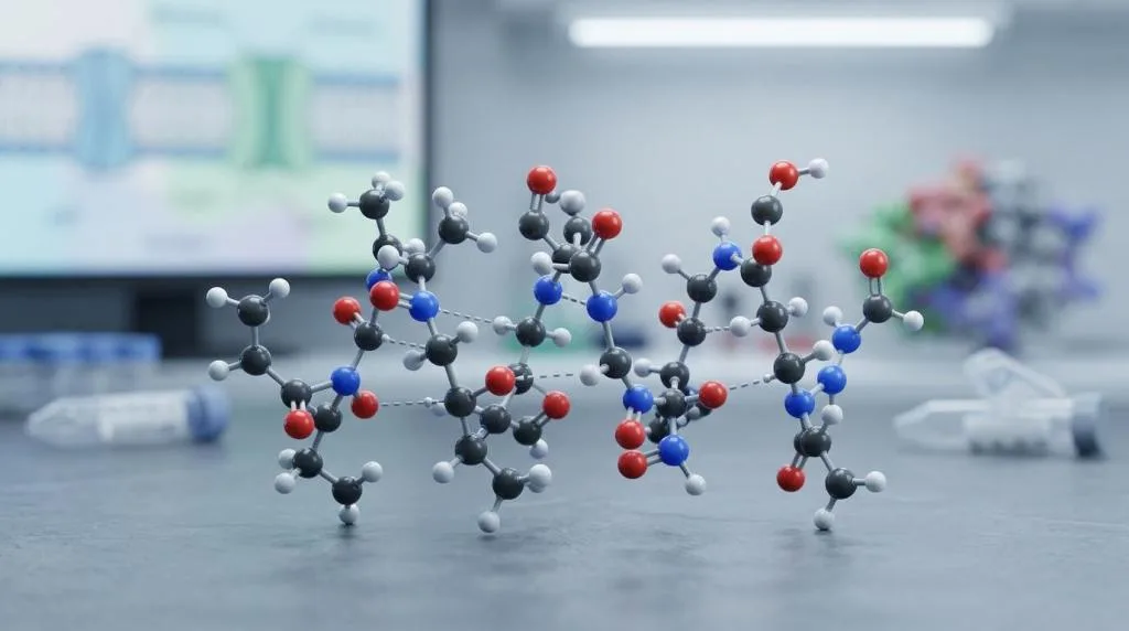
Peptides Explained: How Sequence Drives Function in Cells
Peptides Explained: How Sequence Drives Function in Cells details how amino-acid order controls structure, binding, stability, and cellular activity in research-grade peptide studies.
📋 Table of Contents
Peptides Explained: How Sequence Drives Function in Cells means that a peptide’s amino-acid order—not just its chemical composition—determines the shapes it can adopt, the targets it can bind, and the cellular responses it can trigger. In research, a short chain of up to about 50 residues can act as a hormone signal, receptor ligand, antimicrobial agent, or trafficking cue because the sequence dictates structure, and structure dictates interaction. This sequence-to-structure-to-function link is the core mechanism researchers study when optimizing peptides for cell biology, drug discovery, and assay development.
Key takeaways
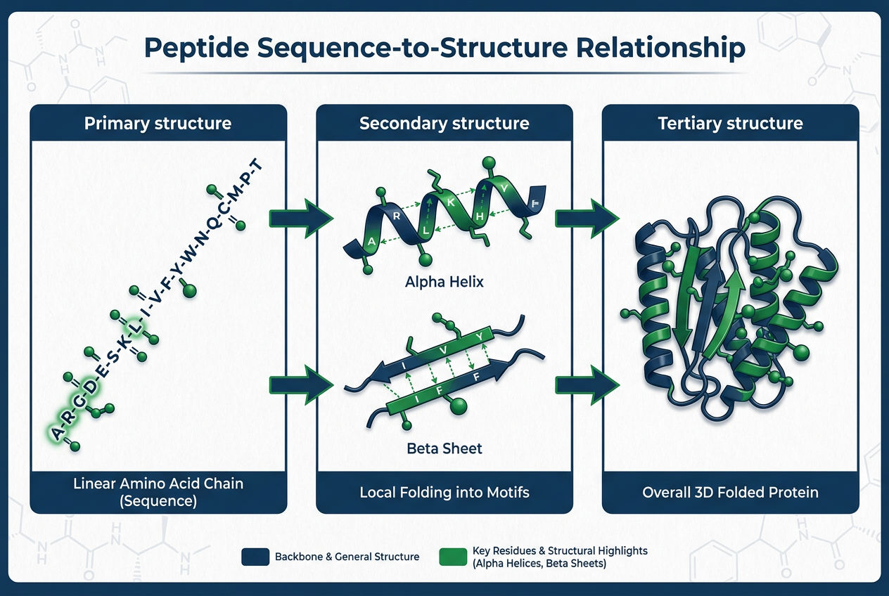
- Peptides Explained: How Sequence Drives Function in Cells is a research framework: sequence → structure → binding → biological response.
- A single residue change can shift charge, remove a hydrogen bond, break a helix, or alter receptor affinity by orders of magnitude.
- Sequence features such as charge distribution, hydrophobic placement, glycine/proline frequency, and cysteine positions control folding and degradation.
- Peptide drug design balances binding, stability, selectivity, exposure, and manufacturability through sequence edits and chemical modifications.
- Screening methods such as phage display and alanine scanning reveal which residues drive activity.
What Do Peptides Do in Cells?
Cells use peptides as information-rich molecules. Some serve as signals released into the extracellular space. Others act locally at membranes, inside vesicles, or in the cytosol. Many work by binding receptors, enzymes, transporters, or nucleic acids with enough selectivity to trigger a defined response. This is why peptides matter far beyond a basic biochemistry lesson. A short sequence can tell a receptor to switch on, help a molecule cross or associate with a membrane, or mark a protein region for interaction. The FDA has also noted that peptide drug candidates often show features of both small molecules and biologics, which reflects how broad their behavior can be.
Peptides sit in a sweet spot between small molecules and full-length proteins. They are usually short chains of amino acids, often up to about 50 residues, yet they can act with striking precision inside cells. Hormone signaling, receptor activation, antimicrobial defense, trafficking cues, and molecular recognition can all depend on peptides whose activity comes down to the order of a few residues. That central idea is simple and powerful: sequence drives function. A peptide’s amino-acid sequence limits the shapes it can adopt, those shapes affect what it can bind, and binding patterns shape the biological response. Current cell biology, structural biology, and drug-development work all point back to that sequence-to-structure-to-function link. This is the essence of Peptides Explained: How Sequence Drives Function in Cells as a research principle.
Why Is Peptides Explained: How Sequence Drives Function in Cells a Core Research Framework?
The sequence is often called the primary structure, and that phrase is more than bookkeeping. Each residue brings size, charge, polarity, hydrogen-bonding potential, and conformational preferences. Put those residues in one order and a peptide may fold into a stable helix. Rearrange them slightly and the same chemical parts may become flexible, exposed to solvent, or unable to bind the same target. A 2024 review in Nature Reviews Molecular Cell Biology emphasized that design methods have improved because researchers now have better rules linking sequence, structure, and function. That matters for natural peptides and engineered ones alike. It also helps explain why sequence comparisons can suggest function long before a peptide is tested in a cell.
Several sequence features tend to matter early when scientists evaluate peptide behavior. Charge distribution, hydrophobic residue placement, glycine and proline frequency, aromatic side-chain patterning, cysteine positions, net length, and terminal chemistry all contribute. No single feature gives the whole answer. Cells respond to the combination. Peptides Explained: How Sequence Drives Function in Cells therefore requires a systems view, not a one-residue explanation. Researchers working with peptide libraries often use these features to rank candidates before functional assays. For example, Peptide Libraries: 7 Essential Research Formats for 2025 shows how sequence diversity is structured to probe these variables efficiently.
💡 Key Takeaway: Why Is Peptides Explained: How Sequence Drives Function in Cells a Core Research Framework?
How Does Peptide Sequence Shape Structure Inside Cells?
A peptide does not float through the cell as a simple string. Its backbone twists, side chains pack, hydrogen bonds form or fail to form, and hydrophobic segments may collapse inward or toward a membrane surface. Even when a peptide is dynamic rather than rigid, its sequence still sets the boundaries of that motion. This is where structural motifs become useful. Some sequences favor alpha-helical segments. Others promote turns, beta-rich assemblies, or highly disordered ensembles. NCBI’s cell biology references have long pointed out that amino-acid sequence can predict structural elements, including membrane-spanning alpha helices in larger proteins. The same logic applies to peptide segments: sequence biases structure, and structure biases interaction.
The table below captures common sequence features and the kinds of effects they can create in cells.
| Sequence feature | Structural tendency | Likely cellular effect |
|—|—|—|
| Clustered hydrophobic residues | Burial, self-association, membrane affinity | Stronger membrane interaction, altered uptake, aggregation risk |
| Alternating polar and nonpolar residues | Amphipathic surfaces | Receptor contact or membrane binding with directional selectivity |
| Proline in a helical region | Helix disruption, kink formation | Reduced receptor fit or changed conformational switching |
| Glycine-rich segments | Higher flexibility | Broader conformational sampling, weaker binding in some cases |
| Cysteine pairing potential | Disulfide formation | Greater rigidity and protease resistance in suitable environments |
| Positive charge enrichment | Electrostatic attraction to negative surfaces | Binding to membranes, nucleic acids, or acidic protein patches |
| Cyclized backbone or side-chain link | Restricted motion | Better stability, altered selectivity, longer residence time |
A short sequence, then, can encode far more than composition alone. It can encode shape preference, dynamics, exposure of key residues, and resistance to degradation all at once. This structural encoding is a recurring theme in Peptides Explained: How Sequence Drives Function in Cells. Researchers designing cell-penetrating peptides rely on these rules to balance membrane interaction with cytosolic access. Cell Penetrating Peptides for Research | Peptides Library provides a practical view of how sequence features such as arginine content and amphipathicity influence uptake in laboratory models.
How Does Peptide Binding Depend on Sequence and Shape?
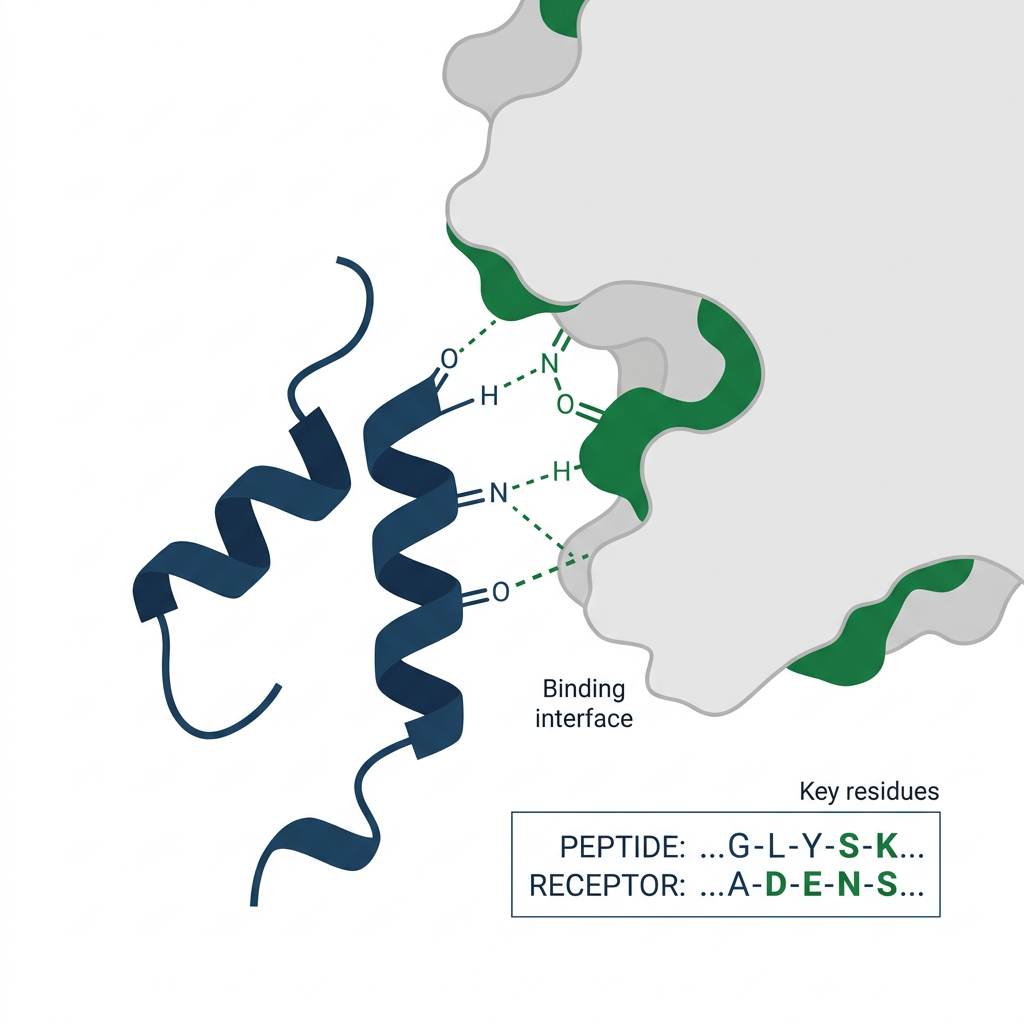
Cellular function appears when a peptide meets a target. Receptors do not “read” a peptide like text. They sense a three-dimensional arrangement of atoms, local charge, hydrophobic surfaces, and hydrogen-bond donors or acceptors presented at the right distances. Sequence matters because it determines whether that arrangement appears often enough, long enough, and in the right cellular context. A peptide that binds a receptor with high affinity usually presents a recognizable motif while minimizing unfavorable contacts. One residue may anchor into a hydrophobic pocket. Another may form a salt bridge. A third may stabilize the backbone in a binding-ready conformation. If any of those pieces shift, signaling can weaken, strengthen, or change direction.
This is also why sequence-guided design has become so important in drug research. Recent work across protein and peptide engineering uses sequence-based and structure-based calculations, often paired with machine learning, to improve target interaction and selectivity. The goal is not only stronger binding. It is better binding to the right target, in the right place, with fewer off-target effects. Peptides Explained: How Sequence Drives Function in Cells highlights that binding is a three-dimensional recognition event, not a linear code. Even when two peptides share similar composition, different residue order can present different surfaces to the same receptor. High-throughput peptide libraries help map this relationship by testing thousands of sequence variants in parallel. Peptide Research Library: A Complete Reference Guide outlines how such libraries are constructed and screened for binding activity.
Why Can a Single Amino-Acid Change Alter Biological Activity?
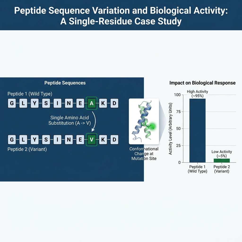
Sometimes one substitution is enough to change everything. A single amino-acid change can alter charge, remove a hydrogen bond, add steric bulk, reduce flexibility, or break a helix. In a receptor-binding peptide, that may reduce affinity by orders of magnitude. In a cell-penetrating or membrane-active peptide, it may change localization, toxicity, or uptake. In a signaling peptide, it may bias one pathway over another. This sensitivity is not surprising when the active surface is small. Large proteins can sometimes absorb local changes because they have many contact points and stabilizing interactions. Short peptides often have less room for error. Their function may depend on a handful of residues arranged just right. That small scale is one reason peptide science remains so exciting. It gives researchers the chance to tune biology with precise edits, then read out the results clearly.
Peptides Explained: How Sequence Drives Function in Cells becomes especially vivid in structure-activity relationship studies. A single alanine substitution can reveal whether a side chain contributes to binding, folding, or stability. A single D-amino acid replacement can test conformational requirements. A single proline insertion can probe helix disruption. These experiments are routine in peptide research and drug development because the biological output is often binary or graded in a predictable way. For laboratories building focused libraries, understanding which positions tolerate substitution is critical. Peptide Library: Research-Grade Peptides for Laboratories discusses how positional scanning and truncation libraries support this type of sequence-function mapping.
💡 Key Takeaway: Why Can a Single Amino-Acid Change Alter Biological Activity?
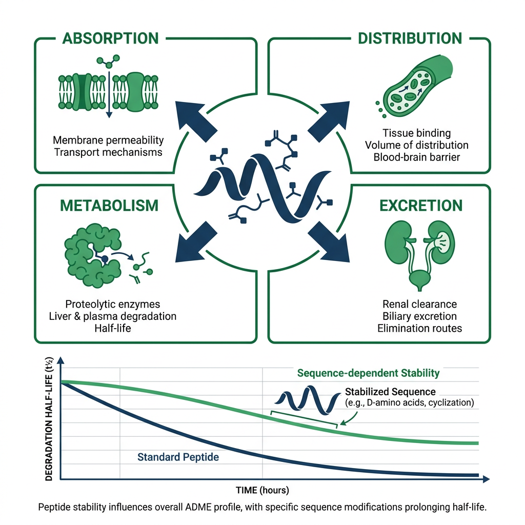
How Does Sequence Affect Stability and ADME in Peptide Drug Design?
Cellular activity is only part of the story. A peptide can bind beautifully in a test tube and still fail in practice if it is unstable, rapidly cleared, or poorly distributed. Drug-development groups pay close attention to ADME properties, meaning absorption, distribution, metabolism, and excretion. Sequence affects all four. Proteases are a major pressure point. Exposed cleavage motifs, flexible backbones, and unprotected termini can shorten peptide lifetime. Chemical and structural changes are often used to address that problem. FDA materials on peptide drug development point to approaches like cyclization, pseudo-peptide bonds, unnatural amino acids, and peptide conjugation as ways to improve pharmacokinetic behavior. Those changes work because they alter what the sequence “means” in structural terms. Restrict the backbone and a protease may no longer recognize it. Change a residue to an unnatural analog and receptor affinity may stay high while metabolism slows. Add a lipid or polymer conjugate and tissue distribution can shift.
When scientists evaluate sequence changes for therapeutic use, they are usually balancing several goals at once. Binding requires preserving the residues that make the key target contacts. Stability requires reducing protease-sensitive motifs and excessive flexibility. Selectivity requires avoiding motifs that encourage unwanted target interactions. Exposure requires improving circulation time or tissue access when needed. Manufacturability requires keeping synthesis and purification practical. That balancing act explains why peptide optimization rarely stops after the first active sequence is found. Peptides Explained: How Sequence Drives Function in Cells applies to ADME just as much as to target binding, because sequence edits change degradation, clearance, and distribution in measurable ways.
| Optimization goal | Sequence-level consideration | Common research strategy |
|—|—|—|
| Binding | Preserve key contact residues | Alanine scanning, positional libraries |
| Stability | Remove protease cleavage motifs | Cyclization, D-amino acids, N-methylation |
| Selectivity | Avoid promiscuous binding motifs | Negative screening, counter-target assays |
| Exposure | Improve circulation or tissue access | Lipid conjugation, PEGylation, albumin binding |
| Manufacturability | Reduce aggregation and difficult couplings | Sequence redesign, shorter analogs, linker choice |
How Do Researchers Test Sequence-Function Relationships in Peptides?
Much of peptide science moves forward by systematic variation. Change one residue. Shift a charge. Introduce cyclization. Replace a natural amino acid with a close analog. Then measure what happens to structure, binding, signaling, trafficking, and degradation. Step by step, the rules become clearer. Phage display has been especially valuable in this area because it can screen very large peptide collections for binding to a target, then recover the sequence of hits. Alanine scanning, truncation series, and positional scanning libraries provide complementary information about which residues are essential. Peptides Explained: How Sequence Drives Function in Cells is therefore an empirical discipline as much as a theoretical one.
Modern screening workflows often combine several methods. A phage display campaign may identify initial hits. Next, alanine scanning narrows the minimal active sequence. Then structural methods such as NMR, X-ray crystallography, or cryo-EM reveal how the peptide contacts its target. Finally, cell-based assays measure downstream signaling, trafficking, or antimicrobial activity. Throughout this process, sequence is the common variable. Every round of design and testing refines the sequence-function map. Research-grade peptide libraries make this iterative process more efficient because they allow parallel evaluation of many sequence variants. For example, Peptide Libraries: 7 Essential Research Formats for 2025 describes formats such as one-bead-one-compound, phage display, and SPOT synthesis that accelerate sequence-function discovery.
What Does Peptides Explained: How Sequence Drives Function in Cells Mean for Modern Screening?
Modern peptide discovery increasingly relies on large sequence spaces and machine learning to predict activity before synthesis. Sequence-based models can score peptides for membrane binding, protease resistance, and receptor affinity. Structure-based models can filter out sequences unlikely to adopt a binding-competent conformation. These tools do not replace experiments, but they reduce the number of candidates that must be made and tested. Peptides Explained: How Sequence Drives Function in Cells provides the conceptual foundation for these models. A model is only as good as the sequence-structure-function rules it learns, and those rules come from systematic experiments.
Cell-based screening also benefits from sequence-focused design. When a library is built around a known motif, even small variations can reveal how each position contributes to activity. When a library is built randomly, hits may point to entirely new sequence motifs. Both approaches are valuable, and both depend on the principle that sequence drives function. For laboratories designing cell-penetrating peptides, antimicrobial peptides, or receptor ligands, sequence diversity is not noise—it is the primary experimental variable. Cell Penetrating Peptides for Research | Peptides Library illustrates how sequence changes affect uptake, endosomal escape, and cytosolic delivery in research settings.
💡 Key Takeaway: What Does Peptides Explained: How Sequence Drives Function in Cells Mean for Modern Screening?
Why Does Peptides Explained: How Sequence Drives Function in Cells Matter for Peptide Stability?
Stability is a sequence-dependent property, not a separate add-on. A peptide with a flexible glycine-rich loop may be more susceptible to proteolysis than a cyclized analog with disulfide constraints. A peptide with exposed hydrophobic patches may aggregate in serum or bind nonspecifically to membranes. A peptide with a free N-terminus may be degraded by aminopeptidases faster than one with an acetylated or lipidated terminus. Each of these outcomes is encoded in the sequence and can be modified by chemical design. Peptides Explained: How Sequence Drives Function in Cells therefore includes stability as a readout of sequence, alongside binding and signaling.
Researchers often assess stability in parallel with activity. A peptide may show excellent receptor binding but a half-life of minutes in serum. By mapping which residues are cleaved, researchers can introduce targeted modifications that preserve activity while extending lifetime. Cyclization, pseudo-peptide bonds, unnatural amino acids, and conjugation are all sequence-level or structure-level interventions. They change the local chemistry or backbone flexibility that proteases recognize. The FDA has highlighted these strategies in peptide drug development because they address the most common failure point for peptide candidates: rapid metabolism. PubMed and FDA provide additional regulatory and scientific context for peptide stability and pharmacokinetic optimization.
How Does Sequence Influence Membrane Interaction and Cellular Uptake?
Many research peptides act at or across membranes. Antimicrobial peptides often use cationic and hydrophobic residues to disrupt microbial membranes. Cell-penetrating peptides often use arginine-rich sequences to engage cell-surface proteoglycans and trigger uptake. Membrane-active peptides may form amphipathic helices that align at the lipid interface. In every case, sequence determines the balance of charge, hydrophobicity, and secondary structure that governs membrane interaction. Peptides Explained: How Sequence Drives Function in Cells is especially clear in this context because small sequence changes can switch a peptide from inert to membrane-active or from selective to toxic.
For example, increasing positive charge may improve binding to negatively charged membranes but also increase nonspecific toxicity. Adding hydrophobic residues may deepen membrane insertion but reduce solubility. Introducing a proline may break a helix and lower membrane activity. These trade-offs are routinely explored in peptide libraries designed for antimicrobial or cell-penetrating research. Cell Penetrating Peptides for Research | Peptides Library discusses how sequence features such as arginine content, tryptophan placement, and backbone cyclization affect uptake efficiency in laboratory models. The same sequence-function logic applies to peptide hormones and receptor ligands, where membrane proximity can influence receptor engagement.
How Do Sequence Comparisons Predict Peptide Function Before Testing?
Sequence comparison can suggest function long before a peptide is tested in a cell. Conserved residues across related peptides often indicate positions required for structure or activity. Variable positions may tolerate substitution without losing function. Motifs such as signal sequences, nuclear localization signals, and antimicrobial patterns can be recognized from sequence alone. This predictive power is a practical benefit of Peptides Explained: How Sequence Drives Function in Cells. It allows researchers to prioritize candidates, design focused libraries, and avoid wasting resources on sequences unlikely to be active.
Bioinformatics tools search for known motifs, predict secondary structure, and estimate physicochemical properties such as isoelectric point, hydrophobicity, and instability index. These predictions are not perfect, but they narrow the search space. A 2024 review in Nature Reviews Molecular Cell Biology emphasized that improved design methods now link sequence, structure, and function more reliably. When combined with experimental validation, sequence-based prediction accelerates discovery. Research-grade peptide libraries often incorporate such predictions into library design. Peptide Research Library: A Complete Reference Guide explains how sequence diversity is organized to test these predictions systematically.
💡 Key Takeaway: How Do Sequence Comparisons Predict Peptide Function Before Testing?
What Are the Most Common Sequence Motifs in Functional Peptides?
Functional peptides often contain recognizable motifs that recur across biological systems. Signal peptides direct secretion or membrane insertion. Nuclear localization signals contain clusters of basic residues. Antimicrobial peptides frequently alternate hydrophobic and cationic residues to form amphipathic structures. Cell-penetrating peptides often contain multiple arginines. Receptor-binding peptides may present aromatic residues for hydrophobic pockets and acidic or basic residues for salt bridges. Peptides Explained: How Sequence Drives Function in Cells helps researchers recognize these motifs and understand why they work.
Motif recognition is not just a lookup exercise. The same motif can behave differently depending on flanking residues, overall length, and terminal chemistry. A basic cluster may be a nuclear localization signal in one context and a membrane-binding patch in another. An amphipathic helix may be antimicrobial or receptor-binding depending on the distribution of polar and nonpolar residues. This is why sequence context matters as much as motif presence. Peptide libraries that vary flanking sequences around a core motif are especially useful for mapping context effects. Peptide Library: Research-Grade Peptides for Laboratories provides examples of how such libraries are designed and screened in research laboratories.
Why Is Cyclization a Key Sequence-Driven Strategy in Peptide Research?
Cyclization is one of the most effective ways to change what a sequence means in structural terms. A linear peptide with a flexible backbone may sample many conformations, only a few of which bind the target. Cyclization restricts motion, often stabilizing the bioactive conformation and improving binding affinity. It can also protect against exopeptidases by removing free termini and against endopeptidases by hiding cleavage sites. Disulfide bonds, lactam bridges, and backbone cyclization are common research approaches. Peptides Explained: How Sequence Drives Function in Cells highlights cyclization as a sequence-level intervention because it changes conformational freedom without necessarily changing residue identity.
Cyclized peptides often show improved stability, longer residence time, and better selectivity. However, cyclization can also reduce flexibility needed for induced-fit binding or membrane translocation. Researchers therefore test multiple cyclization positions and linker lengths to find the best balance. This is another area where systematic sequence variation is essential. By comparing linear and cyclized versions of the same sequence, researchers can isolate the contribution of backbone constraint. The FDA has noted that cyclization and related chemical modifications are common in peptide drug candidates because they address pharmacokinetic weaknesses while preserving target engagement.
How Does Sequence Affect Aggregation and Solubility in Research Peptides?
Aggregation and solubility are sequence-dependent properties that can make or break a research peptide. Hydrophobic stretches promote self-association and aggregation, especially at high concentration or in aqueous buffers. Aromatic residues can drive pi-stacking interactions that lead to fibril formation. Charged residues improve solubility but may reduce membrane penetration. Glycine and proline influence backbone flexibility and can disrupt ordered aggregates. Peptides Explained: How Sequence Drives Function in Cells includes these practical considerations because a peptide that aggregates may be inactive or toxic in cell assays even if its intended target interaction is strong.
Researchers often modify sequences to reduce aggregation without losing activity. Replacing a hydrophobic residue with a polar analog, inserting a proline or glycine, adding a solubilizing tag, or changing the net charge can all improve solubility. The trade-off is that these changes may also alter binding or stability. Peptide libraries that vary aggregation-prone positions help identify sequences that remain soluble and active. Peptide Research Library: A Complete Reference Guide discusses how sequence design can balance solubility, stability, and activity in research-grade peptide collections.
💡 Key Takeaway: How Does Sequence Affect Aggregation and Solubility in Research Peptides?
What Role Does Sequence Play in Peptide Selectivity?
Selectivity is the ability to bind one target over another, and it is encoded in sequence. A peptide that binds a receptor with high affinity may also bind related receptors if it presents a conserved motif. Off-target interactions can occur with homologous proteins, abundant serum proteins, or unrelated membrane surfaces. Peptides Explained: How Sequence Drives Function in Cells emphasizes that selectivity is not simply high affinity; it is the right pattern of contacts in the right context. Small sequence changes can improve selectivity by removing contacts that allow off-target binding while preserving contacts required for the desired target.
Negative screening is a common way to assess selectivity. A peptide library is screened against the target and against counter-targets such as homologous receptors or serum proteins. Hits that bind only the target are prioritized. Sequence analysis of selective and nonselective hits reveals which residues contribute to discrimination. This approach is widely used in receptor ligand discovery and antimicrobial peptide optimization. Peptide Libraries: 7 Essential Research Formats for 2025 describes how counter-screening and subtractive libraries support selectivity profiling in research workflows.
How Do Researchers Use Sequence-Function Data to Design Better Peptides?
Design begins with sequence-function data. Once researchers know which residues are essential, which tolerate substitution, and which cause off-target effects, they can design new sequences with improved properties. This may involve combining motifs from different peptides, introducing non-natural amino acids, or constraining the backbone by cyclization. Peptides Explained: How Sequence Drives Function in Cells provides the rules that guide these design decisions. Without a clear sequence-function map, design becomes guesswork. With it, design becomes a rational process that can be iterated quickly.
Machine learning has accelerated this cycle. Models trained on large sequence-activity datasets can predict activity for new sequences, rank candidates, and suggest substitutions that improve stability or selectivity. These models rely on the same sequence-to-structure-to-function principle that underlies Peptides Explained: How Sequence Drives Function in Cells. They learn patterns that may be too complex for simple rules, but they still require high-quality experimental data. Research laboratories increasingly combine library screening, biophysical assays, and machine learning to design peptides with desired cellular functions. Peptide Library: Research-Grade Peptides for Laboratories offers a starting point for laboratories building the sequence diversity needed for such data-driven design.
Why Is Sequence Context More Important Than Individual Residues?
A residue does not act in isolation. Its contribution depends on neighboring residues, overall fold, terminal modifications, and the cellular environment. A lysine may be a key salt-bridge partner in one sequence and a nonspecific membrane anchor in another. A tryptophan may sit in a hydrophobic pocket or drive aggregation depending on flanking residues. Peptides Explained: How Sequence Drives Function in Cells therefore treats sequence as a system, not a list of independent parts. This is why single-residue substitutions can have unpredictable effects unless the surrounding context is considered.
Context effects are especially important in short peptides, where the active surface is small and every residue contributes. A substitution that seems conservative—such as replacing leucine with isoleucine—may still alter packing enough to reduce affinity. Researchers use positional scanning, alanine scanning, and combinatorial libraries to map context effects. By varying one position while holding others constant, they can measure the contribution of each residue in its native context. This systematic approach is central to modern peptide science and to any practical application of Peptides Explained: How Sequence Drives Function in Cells.
💡 Key Takeaway: Why Is Sequence Context More Important Than Individual Residues?
How Does Sequence Influence Peptide Trafficking and Localization?
Cellular trafficking and localization are sequence-encoded. Signal peptides direct nascent proteins to the secretory pathway. Nuclear localization signals direct peptides into the nucleus. Mitochondrial targeting sequences carry peptides to mitochondria. Endosomal escape motifs help cell-penetrating peptides reach the cytosol. Each of these signals is a short sequence pattern recognized by cellular machinery. Peptides Explained: How Sequence Drives Function in Cells extends beyond binding to include these spatial and temporal control mechanisms. A peptide that binds its target in vitro may still fail in cells if it cannot reach the right compartment.
Researchers studying intracellular targets must consider trafficking sequences. A cell-penetrating peptide may enter cells but remain trapped in endosomes unless it contains an endosomal escape motif. A peptide intended for nuclear delivery may need a nuclear localization signal. These motifs can be added to a research peptide without necessarily disrupting target binding, but sequence context matters. Adding a highly basic sequence may change membrane interactions or solubility. Cell Penetrating Peptides for Research | Peptides Library explores how sequence features influence uptake, endosomal release, and cytosolic delivery in laboratory settings.
What Are the Limitations of Sequence-Based Prediction?
Sequence-based prediction has limits. Cellular conditions are crowded, dynamic, and full of competing interactions. A peptide may fold differently in buffer than in the cytosol. It may bind serum proteins, interact with membranes nonspecifically, or be modified by cellular enzymes. Post-translational modifications can change charge, hydrophobicity, and conformation. Peptides Explained: How Sequence Drives Function in Cells acknowledges that sequence is necessary but not sufficient to predict every cellular outcome. Experimental validation remains essential.
Predictions are most reliable for well-characterized motifs and simple biophysical properties such as charge, hydrophobicity, and secondary structure propensity. They are less reliable for complex cellular behaviors such as endosomal escape, immune recognition, or tissue distribution. This is why researchers use sequence predictions to prioritize candidates, not to replace experiments. High-throughput screening, cell-based assays, and in vivo studies are still required to confirm function. The combination of prediction and validation is the most efficient path. Peptide Research Library: A Complete Reference Guide discusses how predictive design and experimental screening are integrated in modern peptide research.
How Does Peptides Explained: How Sequence Drives Function in Cells Apply to Antimicrobial Peptides?
Antimicrobial peptides are a classic example of sequence-driven function. Many are short, cationic, and amphipathic, allowing them to bind microbial membranes and disrupt them. The balance of hydrophobic and basic residues determines selectivity for microbial membranes over mammalian membranes. Too much hydrophobicity can increase hemolytic activity. Too little can reduce antimicrobial potency. Peptides Explained: How Sequence Drives Function in Cells is therefore directly relevant to antimicrobial peptide design, where small sequence changes can shift the therapeutic window.
Researchers use sequence libraries to map the relationship between hydrophobicity, charge, and antimicrobial activity. They measure minimal inhibitory concentration, hemolysis, and membrane permeabilization for each variant. The resulting structure-activity relationships guide the design of peptides with improved selectivity and stability. Cyclization and D-amino acid substitution are often used to improve protease resistance without losing activity. Peptide Libraries: 7 Essential Research Formats for 2025 describes library formats that support antimicrobial peptide discovery and optimization in research settings.
💡 Key Takeaway: How Does Peptides Explained: How Sequence Drives Function in Cells Apply to Antimicrobial Peptides?
What Are the Key Sequence Features for Cell-Penetrating Peptides?
Cell-penetrating peptides often share sequence features such as high arginine content, amphipathicity, or specific motifs like penetratin or TAT. Arginine-rich sequences engage cell-surface proteoglycans and promote uptake. Hydrophobic residues can enhance membrane interaction but may also increase toxicity. Proline-rich motifs can influence intracellular trafficking. Peptides Explained: How Sequence Drives Function in Cells helps researchers understand why certain sequence patterns promote cellular entry and how to modify them for specific research goals.
For example, increasing the number of arginines often improves uptake but may alter endosomal escape. Adding tryptophan can improve membrane affinity but also increase aggregation. Introducing a cyclic constraint can stabilize the active conformation and improve protease resistance. Researchers test these variables systematically using cell-penetrating peptide libraries. Cell Penetrating Peptides for Research | Peptides Library provides a detailed research-oriented discussion of sequence features, uptake mechanisms, and assay considerations for laboratory studies.
Why Does Sequence Affect Receptor Signaling Bias?
Receptor signaling is not always on or off. Some peptides bias a receptor toward one downstream pathway over another. This is called biased signaling or functional selectivity. Sequence determines which receptor conformations a peptide stabilizes, and different conformations can couple to different intracellular effectors. Peptides Explained: How Sequence Drives Function in Cells therefore includes signaling bias as a sequence-dependent outcome. A peptide that binds the same receptor as another may produce a different cellular response because it stabilizes a different active state.
Researchers study signaling bias by measuring multiple downstream readouts, such as G protein activation, beta-arrestin recruitment, or second messenger production. By varying sequence, they can identify residues that favor one pathway over another. This has implications for peptide drug design, where pathway selectivity may improve efficacy or reduce side effects. Sequence libraries and structural studies together reveal how specific residue contacts drive biased signaling. Peptide Research Library: A Complete Reference Guide discusses how such functional diversity is explored with research-grade peptide collections.
How Does Sequence Influence Peptide Immunogenicity?
Immunogenicity is the tendency of a peptide to provoke an immune response, and it is partly sequence-dependent. Certain sequence patterns are more likely to be presented by MHC molecules or recognized by antibodies. Aggregation-prone sequences can enhance immunogenicity by forming repetitive structures. Foreign sequences are generally more immunogenic than self sequences. Peptides Explained: How Sequence Drives Function in Cells includes immunogenicity as a practical consideration for peptide therapeutics and research reagents. A peptide that is highly active may still be unsuitable if it triggers unwanted immune responses.
Researchers can reduce immunogenicity by modifying residues that contribute to MHC binding or antibody recognition, by reducing aggregation, or by introducing tolerizing sequences. However, these changes must not disrupt the desired function. This is another example of the balancing act central to peptide optimization. Sequence-based prediction of immunogenicity is improving, but experimental validation in relevant models remains necessary. The FDA and PubMed provide guidance and literature on immunogenicity assessment for peptide-based products.
💡 Key Takeaway: How Does Sequence Influence Peptide Immunogenicity?
What Is the Role of Terminal Modifications in Sequence Function?
Terminal modifications can change peptide function without changing the internal sequence. N-terminal acetylation, C-terminal amidation, lipidation, and PEGylation are common research modifications. Acetylation and amidation can improve stability by protecting against exopeptidases. Lipidation can improve membrane association and circulation time. PEGylation can reduce renal clearance and immunogenicity. Peptides Explained: How Sequence Drives Function in Cells includes terminal chemistry as part of the sequence-function equation because terminals are part of the primary structure that cells and enzymes encounter.
Terminal modifications can also affect binding. A free N-terminus may be required for receptor contact in some peptides, while an acetylated N-terminus may improve binding in others. C-terminal amidation is often important for peptide hormones. Researchers test terminal variants alongside internal sequence changes to fully optimize a peptide. This is standard practice in peptide drug development and in the design of research-grade peptide libraries. Peptide Library: Research-Grade Peptides for Laboratories discusses how terminal modifications and sequence variations are incorporated into laboratory peptide collections.
How Does Sequence Affect Peptide Conformation Dynamics?
Peptide conformation is rarely static. Many functional peptides are dynamic, sampling multiple conformations in solution. Sequence sets the boundaries of that motion. Glycine-rich segments increase flexibility. Proline restricts backbone angles. Disulfide bonds lock conformations. Hydrophobic clusters promote collapse. Peptides Explained: How Sequence Drives Function in Cells recognizes that dynamics are part of function, not just noise. A peptide may need flexibility to adapt to its target or rigidity to maintain a binding-competent state.
Researchers use NMR, circular dichroism, and molecular dynamics simulations to study peptide dynamics. These methods reveal which residues are ordered, which are flexible, and how sequence changes affect conformational sampling. This information is valuable for design because it explains why some peptides bind with fast kinetics and others with slow kinetics. It also informs stability optimization. A peptide that is too flexible may be rapidly degraded; one that is too rigid may not adapt to its target. Sequence-guided design aims to find the right balance.
What Are the Advantages of Sequence-Focused Peptide Libraries?
Sequence-focused libraries are built to test specific sequence-function hypotheses. They may vary one position, scan all positions, or randomize a motif while holding flanking residues constant. These libraries generate data that directly reveal which residues drive activity. Peptides Explained: How Sequence Drives Function in Cells is the conceptual basis for such libraries. By controlling sequence diversity, researchers can isolate the contribution of individual residues or motifs.
Compared with fully random libraries, focused libraries are smaller and easier to screen. They also produce results that are easier to interpret. For example, an alanine scanning library identifies essential residues. A positional scanning library identifies preferred residues at each position. A truncation library identifies the minimal active sequence. Each of these designs is a direct application of the principle that sequence drives function. Peptide Libraries: 7 Essential Research Formats for 2025 describes these formats and their use in modern peptide research.
💡 Key Takeaway: What Are the Advantages of Sequence-Focused Peptide Libraries?
How Does Sequence Drive Function in Peptide-Protein Interactions?
Peptide-protein interactions are among the most sequence-sensitive events in biology. A peptide may bind a protein surface, a catalytic site, or an allosteric pocket. The interaction depends on shape complementarity, hydrogen bonding, salt bridges, and hydrophobic contacts. Because peptides are short, a few residues often dominate the interaction. Peptides Explained: How Sequence Drives Function in Cells is therefore essential for understanding and engineering peptide-protein interactions. Changing one anchor residue can abolish binding, while changing a non-contact residue may have little effect.
Researchers map peptide-protein interfaces using alanine scanning, structural biology, and binding assays. They identify hot-spot residues that contribute most to affinity. These hot spots are often conserved across related peptides. Sequence libraries that vary hot-spot positions help optimize affinity and selectivity. This approach is widely used in drug discovery for targets such as protein-protein interaction interfaces, enzymes, and receptors. Peptide Research Library: A Complete Reference Guide provides further detail on how peptide libraries are used to study protein interactions in research.
Why Is Peptides Explained: How Sequence Drives Function in Cells Relevant to Peptide Drug Development?
Peptide drug development depends on the sequence-to-structure-to-function link at every stage. Hit identification uses sequence diversity to find active peptides. Lead optimization uses sequence edits to improve binding, stability, selectivity, and pharmacokinetics. Manufacturing uses sequence design to ensure reproducible synthesis and purification. Regulatory assessment uses sequence characterization to define identity and purity. Peptides Explained: How Sequence Drives Function in Cells is therefore not just an academic concept; it is a practical framework for developing research-grade peptides and peptide-based candidates.
The FDA has recognized that peptide drug candidates share features of small molecules and biologics, which means their development requires attention to both chemical and biological properties. Sequence determines many of these properties. By understanding how sequence drives function, researchers can design peptides with better drug-like properties while preserving biological activity. This is why sequence-function studies remain a priority in academic and industrial laboratories. FDA provides regulatory context for peptide development, and PubMed offers peer-reviewed literature on sequence-function relationships.
FAQ
What is the core principle of Peptides Explained: How Sequence Drives Function in Cells?
The core principle is that a peptide’s amino-acid sequence determines its three-dimensional shape, and that shape determines which cellular targets it can bind and which biological responses it can trigger. Sequence drives structure, structure drives binding, and binding drives function.
Why can a single amino-acid change alter peptide activity?
A single substitution can change charge, remove a hydrogen bond, add steric bulk, break a helix, or reduce flexibility. In short peptides, the active surface is small, so one residue can contribute a large fraction of the binding energy or structural stability. This makes even conservative substitutions potentially disruptive.
How do researchers test which residues are important in a peptide?
Researchers use systematic variation methods such as alanine scanning, truncation series, positional scanning libraries, and phage display. They measure changes in structure, binding, signaling, trafficking, and degradation after each sequence edit. This builds a sequence-function map that identifies essential residues and tolerable positions.
What sequence features affect peptide stability and ADME?
Protease cleavage motifs, flexible backbones, unprotected termini, and aggregation-prone hydrophobic stretches reduce stability. Cyclization, pseudo-peptide bonds, unnatural amino acids, terminal modifications, and conjugation are common strategies to improve ADME properties. Sequence determines which of these modifications are needed and how they affect activity.
Does Peptides Explained: How Sequence Drives Function in Cells apply only to drug development?
No. It applies to any research use of peptides, including cell biology studies, antimicrobial peptide research, cell-penetrating peptide development, receptor signaling assays, and peptide library screening. The sequence-to-structure-to-function link is fundamental to understanding how any short peptide behaves in a cellular context.
Ready to Take the Next Step?
Every product includes lot-specific documentation and third-party purity testing.
Explore Our Research Products →Free shipping on qualifying orders · Batch COA on request