


How Peptide Structure Shapes Stability and Function
Peptide structure shapes stability and function through backbone geometry, hydrogen bonding, and conformational constraint. Explore 5 proven research factors.
📋 Table of Contents
Peptide structure shapes stability and function because the three-dimensional arrangement of backbone atoms, side chains, and secondary structure determines how a peptide binds its target, resists enzymatic breakdown, and behaves in solution. A more defined shape generally improves target recognition and proteolytic stability, while excessive flexibility often reduces both. Researchers therefore evaluate sequence, backbone geometry, hydrogen bonding, and conformational constraint before using any peptide in an assay.
Key takeaways
- Peptide structure shapes stability through primary sequence, backbone planarity, hydrogen bonding, and side-chain constraints.
- A stabilized alpha helix or constrained turn often improves binding and protease resistance compared with highly flexible ensembles.
- Primary sequence writes the folding rules, but backbone geometry enforces which conformations are physically accessible.
- For research use only, peptides with documented purity, storage conditions, and conformational quality checks give more reproducible data.
- Strategic substitutions, cyclization, disulfide engineering, and terminal modifications help preserve active geometry.
What makes peptide structure shapes a critical research variable?
Peptides are often described as short chains of amino acids, but that definition barely captures why they matter. Their behavior is not dictated by composition alone. Peptide structure shapes the order of residues, the geometry of the backbone, the placement of side chains, and the degree to which the chain adopts a stable three-dimensional form.
That structural picture has practical consequences. A peptide that folds into a defined shape is often better at binding its target, more resistant to enzymatic breakdown, and more likely to keep its intended activity. A peptide that remains highly flexible may still be useful, but it usually pays a price in consistency, stability, or selectivity.
In peptide science, structure is the bridge between sequence and outcome. Two peptides can contain similar amino acids and still behave very differently if one forms a stable helix while the other samples many shapes in solution. That difference can influence receptor recognition, membrane interaction, solubility, and susceptibility to proteases.
Classic molecular biology references and modern review articles agree on a central point: amino acid sequence is the first determinant of shape. From there, local backbone interactions and side-chain chemistry guide whether the chain stays extended, forms a turn, adopts an alpha helix, or assembles into a beta-rich arrangement. Even a single substitution can shift that balance.
This is why peptide structure shapes are not an abstract topic reserved for structural biologists. They affect how peptides are designed, tested, stored, and interpreted in research settings. For a broader reference, see the Peptide Research Library: A Complete Reference Guide.
How does primary sequence influence peptide structure shapes and backbone geometry?
The starting point is primary structure, meaning the linear amino acid sequence. Every residue contributes size, charge, hydrophobicity, hydrogen-bonding potential, and steric constraints. A run of alanine and leucine may favor helicity, while proline can interrupt regular backbone patterns because of its rigid ring structure. Glycine, with its high flexibility, often increases local motion.
The peptide backbone itself imposes limits. The peptide bond is planar because of its partial double-bond character, so it does not rotate freely. Rotation occurs mainly around the bonds on either side of that peptide bond. Those permitted rotations, often described by backbone torsion angles, define which conformations are physically accessible.
That means a peptide is never free to adopt every imaginable shape. It moves within a constrained landscape. Sequence determines where that landscape is broad and flexible and where it is narrow and structured. In this way, peptide structure shapes the accessible conformational landscape through primary sequence and backbone geometry.
A useful way to think about it is this: primary structure writes the rules, and backbone geometry enforces them.
| Structural feature | What it means | Effect on stability | Effect on function |
|—|—|—|—|
| Primary sequence | Order of amino acids | Sets intrinsic folding tendency and degradation risk | Defines binding motifs and interaction chemistry |
| Peptide backbone | Repeating amide framework with limited rotation | Restricts accessible conformations | Positions side chains for target recognition |
| Hydrogen bonding | Interactions among backbone amide and carbonyl groups | Supports helices, sheets, and turns | Helps maintain active shapes |
| Side-chain chemistry | Charge, polarity, size, aromaticity | Can stabilize or destabilize folding | Tunes affinity, selectivity, and membrane behavior |
| Structural constraint | Cyclization, stapling, disulfides, residue choice | Often improves proteolytic resistance | Can preserve bioactive conformation |
Because peptide structure shapes are constrained by backbone planarity, researchers can use sequence features to tune folding. Helix-promoting residues, proline-induced kinks, glycine-driven flexibility, charge clustering, and hydrophobic patterning all shift the balance.
Peer-reviewed literature indexed by PubMed describes these backbone constraints in detail; see PubMed peptide structure stability search.
💡 Key Takeaway: How does primary sequence influence peptide structure shapes and backbone geometry?
Which secondary structures contribute most to peptide structure shapes and function?
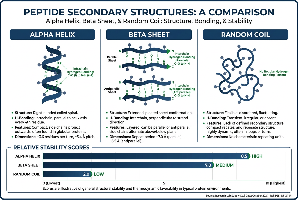
Secondary structure refers to recurring local patterns formed by the peptide backbone. The best known are the alpha helix and beta sheet, both stabilized by hydrogen bonds between backbone amide N-H groups and carbonyl C=O groups. These interactions do not depend primarily on side chains, though side chains strongly affect whether the pattern is favored.
An alpha helix is compact and directional. Side chains project outward from the helical axis, which can create a clear interaction surface. This is valuable when binding depends on residues being displayed at precise spacing. Beta sheets, by contrast, are more extended and can generate broad contact surfaces, though short isolated peptides often struggle to maintain stable sheet structure on their own.
Short peptides frequently lose the structured context they had inside larger proteins. Once isolated, they may no longer hold the turn, helix, or strand arrangement required for activity. Instead, they exist as an ensemble of interconverting conformations, a state often described as conformational heterogeneity.
That is one reason stabilized secondary structure receives so much attention in peptide research. A peptide that spends more time in its biologically relevant shape has a better chance of acting predictably. In practical terms, peptide structure shapes how often the molecule visits active versus inactive conformations.
The following features show how peptide structure shapes folding propensity:
- helix-promoting residues
- proline-induced kinks
- glycine-driven flexibility
- charge clustering
- hydrophobic patterning
In practical design work, researchers often look for these stabilizing elements:
- Backbone hydrogen bonds: support regular helical or sheet-like geometry
- Amphipathic residue spacing: places polar and nonpolar side chains on distinct faces
- Conformational constraints: reduce the number of low-energy alternative shapes
- Strategic substitutions: improve folding tendency without erasing the binding motif
The National Center for Biotechnology Information provides background on peptide and protein structure; see NIH peptide structure resource.
How peptide structure shapes binding surfaces and biological activity
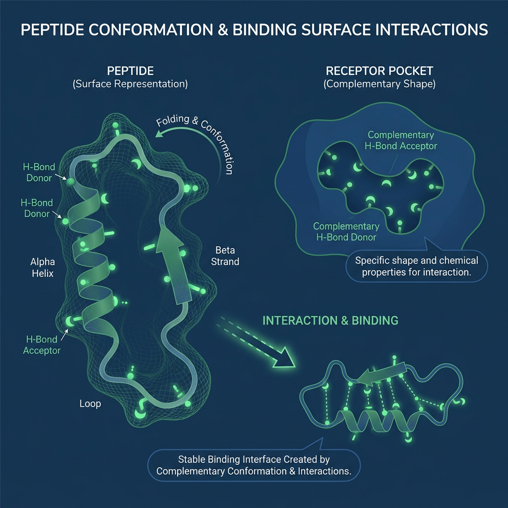
Binding is a geometric event. A target does not recognize a sequence in the abstract. It recognizes a three-dimensional arrangement of atoms, charges, hydrophobic patches, and hydrogen-bond donors or acceptors. If those features are not presented in the right orientation, affinity can drop sharply.
This is why even a small sequence change can have outsized effects. A single amino acid substitution may alter local charge, remove a hydrogen bond, disrupt a helix, or shift side-chain packing. The target may then see a weaker ligand, a different ligand, or no meaningful ligand at all. The relationship between peptide structure shapes and function is often that sensitive.
Selectivity also lives here. A peptide that is too flexible may adapt to multiple surfaces, which sometimes increases off-target interactions. A peptide with a more defined structure can present a cleaner interaction pattern, allowing better discrimination among related receptors or protein surfaces.
One sentence captures the broader lesson: structure does not simply support function, it sets the terms under which function is possible. Thus, peptide structure shapes not only affinity but also selectivity and off-target risk.
For peptides intended for membrane interaction or cellular uptake, Cell Penetrating Peptides for Research | Peptides Library provides additional context on how spatial presentation matters.
What structural design strategies stabilize peptide structure shapes in research?
Researchers use several approaches to increase the probability that a peptide keeps its useful shape in solution or in biological assays. The right approach depends on the peptide length, target class, and intended experimental setting.
Common options include residue substitution, terminal modification, cyclization, disulfide engineering, and helix-stabilizing chemistries. These are not cosmetic changes. They are attempts to tune conformational behavior, exposure of cleavage sites, and the spatial presentation of key side chains.
When reviewing peptide candidates, it helps to ask whether the chosen strategy actually stabilizes the intended peptide structure shapes without erasing the binding motif.
Relevant questions include:
- Does the substitution preserve backbone hydrogen bonding?
- Will cyclization reduce conformational heterogeneity without blocking the target interface?
- Does the terminal modification slow exopeptidase activity?
- Is the disulfide placement compatible with the intended fold?
A Peptide Library: Research-Grade Peptides for Laboratories can support systematic comparison of structural variants.
💡 Key Takeaway: What structural design strategies stabilize peptide structure shapes in research?
How should researchers evaluate peptide structure shapes before assays?
Because peptide structure shapes influence solubility, stability, and binding, researchers benefit from checking measurable attributes before running experiments. Documented purity by HPLC, mass confirmation, and a certificate of analysis (COA) are standard for research-grade peptides.
Storage conditions also matter. Lyophilized peptides generally remain more stable when stored at -20°C or below, protected from moisture and repeated freeze-thaw cycles. Once reconstituted, working aliquots should follow the supplier’s recommended buffer and temperature range.
Conformational quality is harder to measure directly, but circular dichroism or NMR can report on secondary structure content in research settings. These methods show whether peptide structure shapes are consistent with the intended helix, turn, or extended state.
A practical pre-assay checklist includes:
- Confirm identity by mass spectrometry
- Review HPLC purity and COA
- Store lyophilized material at the recommended temperature
- Avoid repeated freeze-thaw of reconstituted aliquots
- Record any visible aggregation or solubility changes
How do flexible and constrained peptide structure shapes compare?
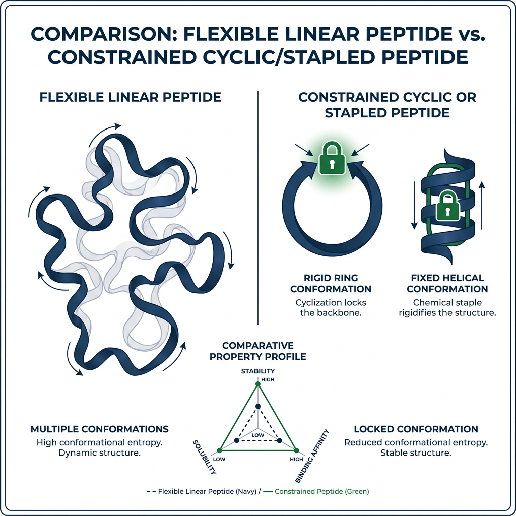
Flexibility is not always a flaw. Some peptides need mobility to engage broad or dynamic targets. Still, excessive flexibility often reduces effective binding because the molecule must pay an energetic cost to adopt the correct shape at the moment of interaction.
That same flexibility can also lower stability in biological environments. Proteases often recognize and cleave peptides that can present an accessible, extended backbone. If a peptide is highly disordered, it may more easily sample cleavage-prone conformations. When the structure is constrained, the enzyme may bind less efficiently.
Helix stabilization, in particular, has been shown to reduce conformational heterogeneity and improve resistance to proteolysis. The logic is straightforward: a more organized backbone can be a poorer substrate for enzymes that prefer flexible, extended chains.
Structural stability and biological stability are often connected, even though they are not identical. This is why direct comparison of flexible and constrained peptide structure shapes is common in research optimization.
| Peptide class | Conformational ensemble | Shape persistence | Typical proteolytic stability |
|—|—|—|—|
| Flexible | Broad | Lower | Often faster enzymatic degradation |
| Constrained | Narrow | Higher | Often improved proteolytic stability |
| Partially folded | Intermediate | Depends on environment, concentration, sequence context | Intermediate |
These patterns show how peptide structure shapes downstream stability in biological assays.
How does peptide structure shapes affect solubility and aggregation in research samples?
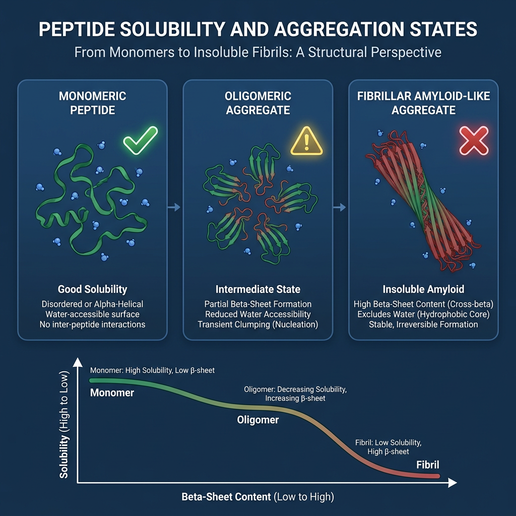
Solubility and aggregation are practical readouts of how peptide structure shapes behave in solution. A peptide that folds into a stable, compact shape may expose fewer hydrophobic patches than the same sequence in a disordered state. Conversely, a flexible peptide can transiently expose hydrophobic side chains, increasing the chance of self-association or precipitation in aqueous buffers.
Researchers often test solubility in the intended assay buffer before committing to a full experiment. If aggregation is observed, structural modifications such as charged residue substitution, terminal capping, or cyclization may reduce self-association. These changes do not alter the core binding motif but can shift peptide structure shapes toward a more soluble ensemble.
This is why solubility screening belongs in the same workflow as purity and mass confirmation. A peptide that is chemically pure but conformationally unstable may still perform poorly. Documenting buffer, pH, temperature, and time in solution helps separate sequence effects from handling effects.
💡 Key Takeaway: How does peptide structure shapes affect solubility and aggregation in research samples?
Which sequence patterns stabilize peptide structure shapes in short peptides?
Short peptides cannot rely on the extensive tertiary contacts that stabilize larger proteins. Instead, local sequence patterns must do more work. Helix-promoting residues such as alanine and leucine can raise helical propensity, while proline often disrupts regular backbone geometry. Glycine increases flexibility and can reduce persistence of any single shape.
Charge clustering and hydrophobic patterning also matter. An amphipathic sequence that places polar and nonpolar residues on opposite faces can stabilize a helix in membrane-mimetic or partially hydrophobic environments. That spatial organization makes peptide structure shapes easier to maintain without large proteins.
Researchers may use these patterns as a design filter. Before synthesis, they can score candidate sequences for helical propensity, proline content, glycine placement, and amphipathic spacing. This step does not guarantee activity, but it narrows the number of variants that must be tested empirically.
What practical checklist supports peptide structure shapes in research workflows?
To keep peptide structure shapes consistent across experiments, researchers can standardize handling and documentation. This reduces variability from lot-to-lot differences, solvent history, and storage conditions.
A recommended workflow includes:
- Use a single batch or lot for comparative studies
- Prepare stock solutions in the same solvent and concentration
- Record storage temperature and time in solution
- Confirm that the COA lists the expected purity and mass
- Run a control peptide with known conformational behavior
This checklist does not replace structural analysis, but it makes peptide structure shapes a controlled variable rather than an uncontrolled one.
Why does peptide structure shapes matter for research-use compliance?
All peptide structure shapes discussed here apply to research use only. No statement in this article implies human consumption, clinical dosing, or therapeutic use. Research peptides should be handled according to institutional guidelines, supplier safety data sheets, and applicable regulations.
When documenting experiments, researchers should note the peptide sequence, purity, storage, and any observed conformational behavior. That documentation supports reproducibility and helps other laboratories compare results. This focus on research-use compliance ensures that peptide structure shapes are evaluated as a scientific variable, not as a product claim.
💡 Key Takeaway: Why does peptide structure shapes matter for research-use compliance?
FAQ
What is the single most important factor in peptide structure shapes?
Primary amino acid sequence is the first determinant, but backbone geometry and side-chain interactions shape the accessible conformations. A single substitution can shift folding, stability, and binding.
Does peptide structure shapes affect proteolytic stability?
Yes. More constrained peptides often present a less accessible backbone to proteases, reducing cleavage. Highly flexible peptides may sample extended conformations that enzymes recognize more easily.
Can a flexible peptide still have useful function?
Some peptides need flexibility to engage dynamic targets. However, excessive flexibility often lowers effective binding because the molecule must pay an energetic cost to adopt the active shape.
How can I verify peptide structure shapes in a research sample?
Use HPLC and mass spectrometry for identity and purity, and circular dichroism or NMR for secondary structure content. Review the COA and follow recommended storage conditions.
Is peptide structure shapes relevant for storage?
Yes. Lyophilized peptides stored at -20°C or below, protected from moisture and freeze-thaw cycles, retain their intended conformation better than poorly stored samples.
Ready to Take the Next Step?
Every product includes lot-specific documentation and third-party purity testing.
Explore Our Research Products →Free shipping on qualifying orders · Batch COA on request