



How Lab Models Evaluate Peptide Activity and Selectivity
Peptide activity and selectivity across lab models: receptor binding, functional assays, structural biology, peptide arrays, and computational prediction for research use only.
📋 Table of Contents
Lab models evaluate peptide activity and selectivity through a tiered system of receptor binding assays, cell-based functional readouts, structural biology, peptide arrays, and computational prediction. No single assay captures the full biological profile of a research peptide, because affinity, efficacy, pathway bias, and off-target engagement can diverge across model systems. This reference explains how each lab model contributes to a defensible peptide activity and selectivity claim for research-use-only peptides.
Key takeaways
- Peptide activity and selectivity cannot be established by one assay; a layered approach using binding, functional, structural, array, and computational models is required.
- Binding assays measure affinity, competition, and residence time, while cell-based assays reveal efficacy, potency, and biased signaling.
- Structural biology and receptor mutagenesis explain the molecular contacts that drive peptide activity and selectivity differences between close analogs.
- Peptide arrays and structure-activity relationship mapping identify the minimal active motif and residues controlling receptor subtype preference.
- Reproducible peptide activity and selectivity data depend on documented purity, storage conditions, and assay controls, all within a research-use-only framework.

Why do peptide activity and selectivity require multiple lab models?
In peptide pharmacology, activity usually refers to what a peptide does after it meets its target. Selectivity refers to how strongly it prefers one target, receptor state, or signaling path over others. Those two properties are related, though they are not the same.
A peptide can show strong affinity in a binding assay and still behave as a weak agonist, a partial agonist, an antagonist, or a ligand with pathway bias. That is especially relevant in GPCR research, where receptor engagement may lead to G protein signaling, β-arrestin recruitment, desensitization, trafficking, or endosomal signaling, depending on the system used.
Researchers usually look across several layers of evidence before calling a peptide active or selective:
- Affinity at the intended target
- Potency in functional assays
- Maximal efficacy
- Pathway preference
- Off-target binding
- Residence time
This layered approach matters because no single readout can fully represent peptide behavior in a living system. A peptide may bind tightly to a receptor yet produce little downstream signaling. Another may appear highly selective in one assay format, then lose that edge when receptor density, temperature, or readout changes. Strong peptide activity and selectivity claims therefore depend on agreement across independent lab models, not on one favorable curve.
How do receptor binding assays quantify peptide affinity and residence time?
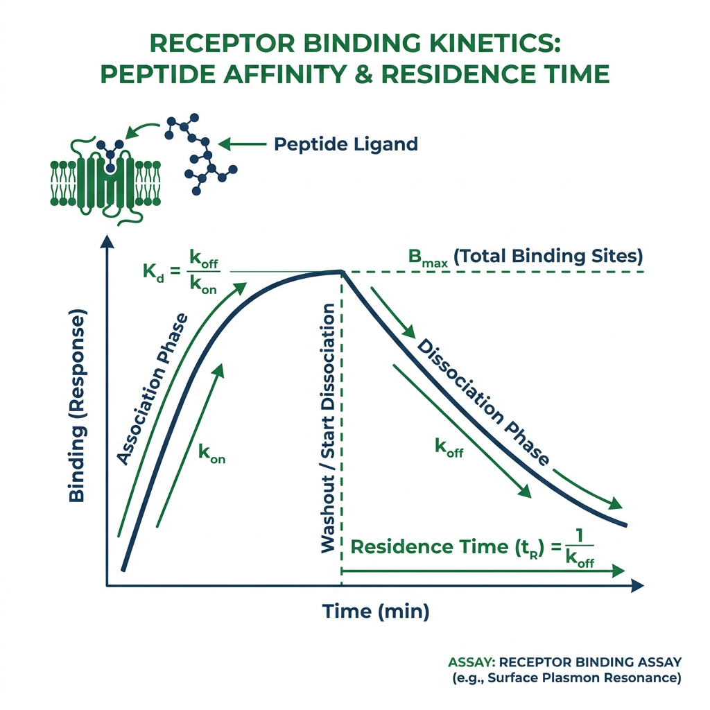
Binding assays are often the first checkpoint. They ask a direct question: does the peptide bind the receptor, and how strongly? In practice, this can mean saturation binding, competition binding, or kinetic studies that estimate association and dissociation rates.
These assays are highly useful for comparing close analogs. A small sequence change, terminal modification, or residue substitution can shift affinity by orders of magnitude. In peptide programs, that makes binding assays a fast way to map structure-activity trends before more complex cellular work begins.
They also help separate occupancy from function. If a peptide fails in a cell assay, the issue may be poor receptor binding, limited stability, low membrane access, or weak coupling to signaling. Binding data narrow that uncertainty.
| Assay format | Typical readout | Main strength | Common limitation |
|—|—|—|—|
| Filtration binding | Bound versus free ligand after separation | Widely used and direct | Wash steps can disturb weak or fast-off interactions |
| Scintillation proximity | Signal from radioligand near bead-bound receptor | Homogeneous format, good sensitivity | Requires specialized reagents and setup |
| Fluorescence polarization | Change in polarization on binding | Non-radioactive and adaptable | Signal quality depends on tracer design and assay window |
| Competition binding | Displacement of a labeled ligand | Efficient for ranking analogs | Interpretation depends on tracer choice and receptor state |
Kinetic parameters add another layer that is easy to miss. A peptide with a slow dissociation rate constant may show longer receptor occupancy and different biological performance than a peptide with the same equilibrium affinity but rapid dissociation. That can influence peptide activity and selectivity claims under non-equilibrium conditions, which are often closer to what tissues experience than an idealized equilibrium assay.
Assay conditions also matter more than many people expect. Buffer composition, temperature, incubation time, receptor preparation, and tracer selection can shift measured affinity. A peptide that looks clean in one membrane binding setup may behave differently in whole-cell binding or when a different receptor construct is used. Documenting these variables is essential for reproducible peptide activity and selectivity data.
💡 Key Takeaway: How do receptor binding assays quantify peptide affinity and residence time?
How do cell-based functional assays reveal peptide activity and selectivity?
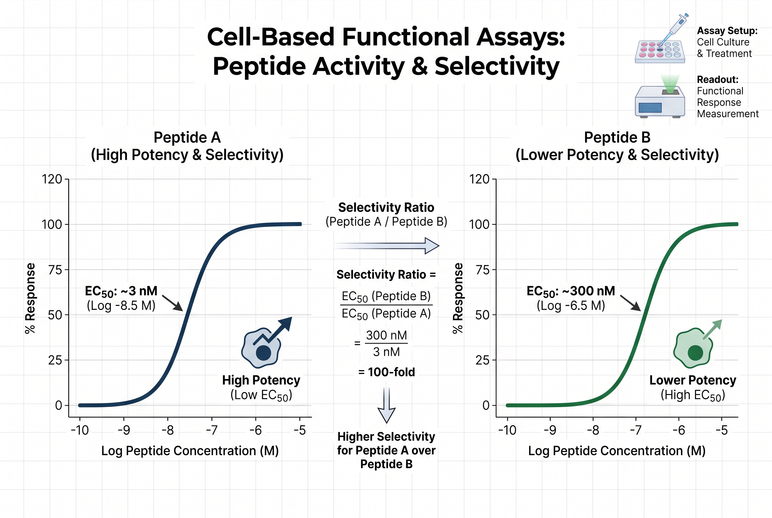
Once binding is established, cell-based assays test whether receptor engagement turns into a biological effect. For peptide ligands acting at GPCRs, these assays commonly measure cAMP, calcium mobilization, IP1 accumulation, ERK phosphorylation, β-arrestin recruitment, receptor internalization, or related downstream events.
This is where efficacy becomes visible. Two peptides can bind the same receptor with similar affinity and still produce very different cellular outcomes. One may act as a full agonist, another as a partial agonist, and another may favor one pathway over another. Current literature often refers to this pathway preference as biased signaling or functional selectivity.
That matters because peptide activity and selectivity is not only about choosing receptor A over receptor B. It can also mean favoring G protein signaling over β-arrestin recruitment, or producing a trafficking profile that changes the timing and location of signaling.
Several cell-based readouts are especially common in peptide research:
- cAMP assays: useful for ranking potency at receptors coupled to Gs or Gi pathways
- Calcium flux assays: sensitive and fast, though signal amplification can exaggerate weak effects
- β-arrestin recruitment assays: valuable for pathway bias, desensitization, and receptor regulation
- Internalization assays: connect activation to trafficking and endosomal behavior
A peptide can look highly potent in one of these systems and modest in another. That does not always mean one assay is wrong. It may reflect receptor reserve, signal amplification, cell background, transducer expression, or true pathway preference. Recent reviews of GPCR assay platforms also point to GRK phosphorylation, β-arrestin-mediated desensitization, and endosomal internalization as useful markers when a simple potency value is not enough.
When comparing peptide activity and selectivity across cell lines, always record receptor expression level, assay incubation time, and whether the readout is proximal or distal to receptor activation. These variables determine whether a potency difference reflects true ligand bias or system-dependent amplification.
How do structural biology and receptor mutagenesis explain peptide selectivity?
Binding and cell data show what happens. Structural work helps explain why.
Crystal structures and cryo-EM models of peptide receptors have made it easier to see how peptides contact extracellular loops, transmembrane residues, and juxtamembrane regions. In class A peptide GPCRs, sequence and mutagenesis work have identified hydrophilic receptor residues that shape preference for peptide terminal chemistry, including C-terminal free carboxyl groups, amidated termini, and N-terminal ammonium features.
For class B GPCRs, classic receptor models and later structural studies support a different theme: peptide engagement of extracellular and juxtamembrane receptor regions helps trigger the receptor conformations that start intracellular signaling. That architecture helps explain why truncation can preserve some binding yet sharply reduce activation.
Receptor mutagenesis is especially valuable when peptide activity and selectivity claims are close. A single residue change can weaken one peptide analog far more than another, showing which contacts drive preference. In practice, mutagenesis often resolves questions that neither a binding curve nor a signaling curve can answer alone.
Structural context also helps interpret puzzling results. A peptide may lose affinity after one substitution, yet keep activity if the changed contact was not essential for the active receptor conformation. Another may keep affinity but lose efficacy if the substitution disrupts the part of the interaction that stabilizes activation rather than mere binding.

How do peptide arrays and SAR mapping refine active motifs?
Before a research group commits to extensive animal or translational work, it usually wants a sharper picture of which residues matter most. Peptide arrays and structure-activity relationship studies are built for that purpose.
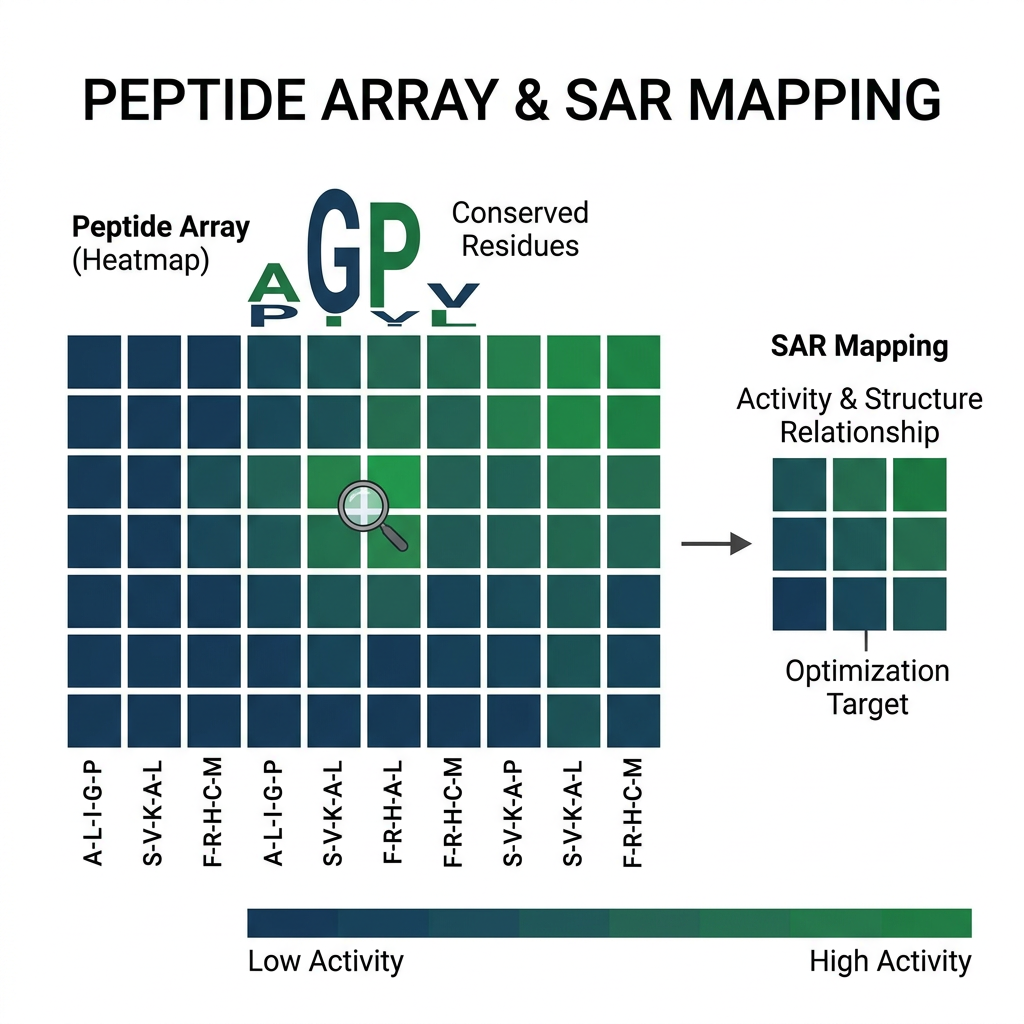
Arrays can screen many short variants in parallel. Truncation series, alanine scans, terminal edits, and non-natural residue substitutions can reveal the minimal active region, tolerated changes, and residues that control peptide activity and selectivity. This is especially helpful when a long peptide has one compact pharmacophore and several positions that mainly tune stability or receptor bias.
Common structure-activity mapping tools include:
- Alanine scanning
- Truncation series
- N-terminal and C-terminal modification
- Charge reversal substitutions
- Cyclization or conformational constraint
- Non-natural residue swaps
These approaches become even more informative when paired with binding and cell-based data. A residue can matter for affinity, signaling bias, protease resistance, or receptor subtype preference, and those effects do not always move together. For example, a substitution that improves metabolic stability may simultaneously reduce selectivity at a closely related receptor, so the two readouts must be tracked together.
A well-designed peptide array can reduce the number of full-length analogs needed for later functional confirmation. When you identify the minimal active motif and the residues that tune peptide activity and selectivity, you can focus synthesis resources on the most informative variants.
💡 Key Takeaway: How do peptide arrays and SAR mapping refine active motifs?
How does computational peptide activity prediction prioritize experiments?
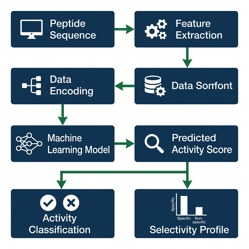
Computational models are now part of many peptide pipelines, especially where receptor structures or large sequence-activity datasets are available. These models can predict binding poses, estimate relative affinity for receptor subtypes, and flag residues likely to influence peptide activity and selectivity before any synthesis begins.
Common computational approaches include molecular docking, molecular dynamics simulations, machine learning models trained on peptide-receptor interaction data, and sequence-based predictors of antimicrobial or receptor-targeting activity. Each method has strengths and limits. Docking works best when a high-resolution receptor structure exists; machine learning can capture patterns from thousands of measured peptide sequences but may fail on novel scaffolds.
Computational prediction does not replace experiments. It prioritizes them. A ranking of candidate peptides by predicted affinity or selectivity can cut the number of binding and functional assays required. When computational output conflicts with experimental peptide activity and selectivity data, the conflict often points to an unmodeled factor such as membrane partitioning, receptor oligomerization, or intracellular trafficking.
For research-use-only peptide programs, computational tools are particularly valuable for screening virtual libraries before committing to custom synthesis. This reduces material use and helps laboratories stay within their peptide research budgets.
What storage conditions and purity metrics support reproducible peptide activity and selectivity data?
Reproducible peptide activity and selectivity data depend on controlling variables outside the assay itself. Two of the most important are peptide purity and storage conditions.
Purity is typically reported as a percentage determined by HPLC, often with mass spectrometry confirmation. For research peptides used in binding or functional assays, a purity of 95% or higher is common, though some exploratory screens tolerate 90–95%. Lower purity can introduce confounding signals from truncated sequences, deletion products, or residual synthesis reagents. Always request a certificate of analysis (COA) that includes HPLC purity, mass identity, and peptide content when available.
Storage conditions directly affect peptide stability. Most lyophilized research peptides should be stored at –20 °C or –80 °C, protected from light and moisture. Once reconstituted, peptides are often aliquoted and stored at –80 °C to avoid repeated freeze-thaw cycles. The exact buffer, pH, and concentration can influence aggregation, oxidation, and degradation, all of which alter measured peptide activity and selectivity. Document storage temperature, reconstitution solvent, and freeze-thaw history for every assay.
A peptide that has degraded may still bind its target but show reduced functional potency or altered selectivity. For example, oxidation of methionine residues or deamidation of asparagine can change receptor contacts without eliminating binding. This is why stability testing under assay conditions is a practical part of peptide activity and selectivity evaluation.
Which assay combinations give the most defensible peptide activity and selectivity profile?
No universal combination fits every peptide, but a defensible profile usually includes at least one binding assay, one cell-based functional assay, and one structural or array-based analysis. For GPCR peptide ligands, a common tiered approach is:
1. Primary screen: competition binding against the intended receptor and at least one closely related off-target receptor.
2. Functional confirmation: cAMP or calcium assay to establish potency and efficacy; β-arrestin recruitment to assess pathway bias.
3. Selectivity panel: binding or functional assays against a panel of related receptors, chosen based on sequence homology or known cross-reactivity.
4. Structural or SAR support: receptor mutagenesis, peptide array mapping, or computational docking to explain the molecular basis of peptide activity and selectivity.
This combination addresses the three questions that matter most: Does the peptide bind? Does binding produce a functional response? Does the peptide prefer the intended target over others under relevant assay conditions?

When resources are limited, start with a competition binding assay and a single functional readout. If those two agree, add a selectivity panel. If they disagree, move to structural or mutagenesis analysis before investing in more cell lines. The goal is not to collect every possible data point, but to collect data that can independently confirm or challenge the peptide activity and selectivity claim.
For further background on building a complete peptide research library and selecting reference standards, see the Peptide Research Library: A Complete Reference Guide. If your peptide must cross a membrane to reach an intracellular target, review Cell Penetrating Peptides for Research | Peptides Library before choosing a functional assay.
💡 Key Takeaway: Which assay combinations give the most defensible peptide activity and selectivity profile?
How do off-target panels strengthen peptide activity and selectivity data?
Selectivity is only as strong as the off-target panel behind it. A peptide that binds the intended receptor with 10 nM affinity may look excellent until tested against a homologous receptor where it binds with 12 nM affinity. The ratio of affinities or potencies, not the absolute value at the primary target, defines peptide activity and selectivity.
A practical off-target panel should include receptors with high sequence homology, receptors from the same class or family, and any receptor previously reported to interact with similar peptide sequences. For peptide GPCR ligands, this often means testing against multiple subtypes of the same receptor family, plus a small set of unrelated GPCRs to check for nonspecific membrane effects.
Functional off-target screens can reveal differences that binding panels miss. A peptide may bind two receptors with similar affinity but activate only one, or activate both through different pathways. Running a functional selectivity panel, even with a single readout, adds biological relevance to peptide activity and selectivity claims.
Why do assay conditions change peptide activity and selectivity measurements?
Assay conditions are not neutral. Receptor density, temperature, buffer composition, incubation time, and tracer concentration all influence measured peptide activity and selectivity. A peptide that appears selective at 25 °C may lose that selectivity at 37 °C because receptor conformation or membrane fluidity changes.
Receptor reserve is another common source of discrepancy. In systems with high receptor reserve, a weak partial agonist can produce a near-maximal response, masking low intrinsic efficacy. In systems with low reserve, the same peptide may look like a weak agonist or antagonist. This does not mean one result is wrong; it means peptide activity and selectivity must be interpreted in the context of the assay system.
Tracer choice in competition binding also matters. A radiolabeled agonist and a radiolabeled antagonist can report different affinity states of the receptor, leading to different rank orders for the same set of peptides. Whenever possible, run competition binding with both an agonist and an antagonist tracer, or use a functional assay to confirm the binding result.

How can peptide arrays be combined with computational docking to refine peptide activity and selectivity?
Peptide arrays generate empirical structure-activity data; computational docking provides a structural hypothesis for that data. Combining the two accelerates the path from a long peptide to a minimal active motif with defined peptide activity and selectivity.
A typical workflow starts with an alanine scan or truncation series on the peptide array. The resulting activity map highlights residues that are essential for binding, residues that tune efficacy, and residues that influence receptor subtype preference. Computational docking then places the peptide in the receptor binding pocket and tests whether the essential residues form the predicted contacts. If docking agrees with the array, confidence in the model rises. If docking disagrees, the model or the array may need refinement.
This combined approach is especially useful for peptide families with multiple closely related receptors. By docking the same peptide into two receptor subtypes, researchers can predict which residues drive subtype selectivity and then test those predictions with targeted substitutions. The result is a more efficient use of synthesis and assay resources while improving the rigor of peptide activity and selectivity claims.
For laboratories building a focused peptide collection for such studies, the Peptide Library: Research-Grade Peptides for Laboratories offers practical guidance on selecting and organizing peptide sets.
💡 Key Takeaway: How can peptide arrays be combined with computational docking to refine peptide activity and selectivity?
What role does peptide purity play in peptide activity and selectivity assays?
Peptide purity directly affects the interpretation of peptide activity and selectivity data. Impurities can act as competitive inhibitors, partial agonists, or allosteric modulators, shifting the apparent potency or selectivity of the main peptide. A peptide reported as 90% pure may contain 10% of a truncated sequence that binds the same receptor with different efficacy, leading to a misleading functional profile.
Always verify purity by HPLC and mass spectrometry before running critical assays. For quantitative binding and functional studies, a purity of 95% or higher is recommended. If a COA is unavailable, request one or run an in-house analytical check. Document the purity of each peptide batch in the same dataset as the assay results, because batch-to-batch purity variation can explain otherwise puzzling changes in peptide activity and selectivity.
Peptide content, which accounts for residual salts and water, is a separate metric from HPLC purity. Two peptide batches can both show 98% HPLC purity but differ in peptide content, affecting the actual concentration in solution. Correcting for peptide content before preparing stock solutions reduces variability in peptide activity and selectivity measurements.
How do residence time and binding kinetics influence peptide activity and selectivity?
Residence time, the reciprocal of the dissociation rate constant, is a kinetic parameter that equilibrium binding assays do not capture. A peptide with a long residence time may remain bound to its receptor after the free ligand concentration drops, producing prolonged signaling or different desensitization patterns. This can alter peptide activity and selectivity in ways that a simple affinity value misses.
For example, two peptides may have identical equilibrium dissociation constants, but one dissociates in seconds while the other remains bound for minutes. Under non-equilibrium conditions, such as pulsatile hormone release or transient ligand exposure, the slow-dissociating peptide may show greater functional effect or broader apparent selectivity. Kinetic binding assays, such as surface plasmon resonance or radioligand association/dissociation experiments, are therefore valuable complements to equilibrium competition binding.
When a peptide shows unusual peptide activity and selectivity in a functional assay but normal equilibrium binding, measure its binding kinetics before concluding that the functional result is an artifact. The discrepancy may reflect a real difference in residence time or receptor state preference.
How can receptor mutagenesis resolve ambiguous peptide activity and selectivity data?
Receptor mutagenesis is a direct experimental test of the molecular contacts that determine peptide activity and selectivity. By changing a single receptor residue and measuring the effect on peptide binding and function, researchers can identify which contacts are essential for affinity, which for efficacy, and which for subtype preference.
A classic design is to mutate a residue predicted to contact the peptide terminal chemistry, then test a series of peptide analogs with different terminal groups. If the mutation selectively weakens one analog but not another, that residue contributes to peptide activity and selectivity. If the mutation affects all analogs equally, the residue may be important for receptor structure rather than ligand discrimination.
Mutagenesis is also useful for validating computational docking models. A docking model may predict that a specific receptor residue forms a hydrogen bond with a peptide side chain. Mutating that residue to remove the hydrogen bond donor or acceptor should reduce binding if the model is correct. This closes the loop between computational prediction and experimental peptide activity and selectivity data.
💡 Key Takeaway: How can receptor mutagenesis resolve ambiguous peptide activity and selectivity data?
What are the most common pitfalls in peptide activity and selectivity assays?
Even experienced laboratories encounter pitfalls that compromise peptide activity and selectivity data. Recognizing these pitfalls in advance saves time and reduces misleading conclusions.
- Ignoring peptide content: Using nominal mass instead of peptide content leads to concentration errors that shift potency curves.
- Running only one functional readout: Pathway bias means a peptide can be a strong G protein agonist but a weak β-arrestin recruiter, or vice versa.
- Testing selectivity against too few off-targets: A selectivity claim is only as strong as the panel behind it.
- Using degraded peptide stocks: Oxidation, deamidation, or aggregation can alter receptor contacts without eliminating binding.
- Ignoring receptor reserve: High receptor reserve can mask weak partial agonism and inflate apparent potency.
- Failing to document assay conditions: Buffer, temperature, and tracer choice can shift measured affinity and selectivity.
Addressing these pitfalls systematically improves the reliability of peptide activity and selectivity claims. When a result seems too clean, check the controls; when a result seems confusing, add a complementary assay rather than repeating the same one.
How do cell penetrating peptides affect functional peptide activity and selectivity assays?
Some research peptides target intracellular proteins or receptors located in endosomal compartments. For these peptides, membrane access becomes a critical variable in functional assays. A peptide may bind its target with high affinity in a biochemical assay but show no activity in a cell-based assay simply because it cannot cross the plasma membrane.
Cell penetrating peptides (CPPs) or peptide conjugates with CPP sequences can deliver cargo into cells, but the CPP itself may alter peptide activity and selectivity by changing solubility, aggregation, or off-target interactions. When using CPP-conjugated peptides, include appropriate controls: unconjugated peptide, scrambled CPP, and a membrane-impermeable analog if available.
For more detail on CPP selection and assay considerations, see Cell Penetrating Peptides for Research | Peptides Library.
What internal controls should accompany peptide activity and selectivity assays?
Internal controls anchor peptide activity and selectivity data to known reference points. Without them, a potency value or selectivity ratio floats without context.
Common internal controls include:
- A reference agonist or antagonist with well-characterized affinity and efficacy at the same receptor.
- A vehicle control to account for solvent effects.
- A positive control peptide known to activate the pathway being measured.
- A negative control peptide with a scrambled sequence or an inactive analog.
- A receptor expression control, such as a ligand known to activate an unrelated receptor, to confirm cell viability and assay function.
These controls allow researchers to normalize results across plates, days, and batches, reducing false conclusions about peptide activity and selectivity. For example, if the reference agonist shows the expected potency but the test peptide does not, the issue is likely peptide-specific rather than assay-wide.
💡 Key Takeaway: What internal controls should accompany peptide activity and selectivity assays?
How can peptide activity and selectivity data be reported transparently?
Transparent reporting makes peptide activity and selectivity data useful to other researchers and to future projects. At minimum, report the following for each assay:
- Peptide identity, purity, and peptide content
- Storage and reconstitution conditions
- Assay type and readout
- Receptor source and expression level
- Incubation time and temperature
- Number of independent experiments and replicates
- Curve fitting model and constraints
- Affinity or potency values with confidence intervals
- Off-target panel composition
- Any deviations from standard protocols
This level of detail allows others to reproduce the work and to compare peptide activity and selectivity across studies. It also protects against overinterpretation when assay conditions differ. Many journals now require this information in supplementary tables, but including it in internal reports is equally valuable.
How do recent reviews frame peptide activity and selectivity in GPCR research?
Recent reviews of GPCR assay platforms emphasize that peptide activity and selectivity cannot be reduced to a single potency number. They highlight the importance of measuring GRK phosphorylation, β-arrestin-mediated desensitization, and endosomal internalization as complementary markers of ligand behavior. These markers capture aspects of receptor regulation that traditional cAMP or calcium assays miss.
The same reviews note that receptor reserve, signal amplification, and transducer expression levels can shift apparent potency and bias. A peptide that appears G protein-biased in one cell line may appear balanced in another. This is not a failure of the assay; it is a property of the system. The solution is to run multiple functional readouts and to interpret peptide activity and selectivity as a profile, not a single value.
External sources such as PubMed and PMC provide peer-reviewed examples of these assay strategies in peptide GPCR research.
How can a peptide research library support systematic peptide activity and selectivity studies?
A well-organized peptide research library allows systematic comparison of analogs under identical conditions. When peptides are stored, documented, and quality-controlled in a central library, assay variability from batch differences or storage history decreases. This makes peptide activity and selectivity comparisons across analogs more reliable.
Key features of a useful peptide research library include:
- Consistent purity and peptide content documentation
- Uniform storage at –80 °C with minimal freeze-thaw cycles
- Aliquoting to avoid repeated thawing of master stocks
- A searchable database of sequence, purity, COA, and assay history
- Reference peptides with known activity for assay calibration
For guidance on building such a library, see the Peptide Research Library: A Complete Reference Guide. A disciplined library reduces the risk that a change in peptide activity and selectivity is actually a storage artifact.
💡 Key Takeaway: How can a peptide research library support systematic peptide activity and selectivity studies?
What is the role of peptide arrays in identifying receptor subtype selectivity?
Peptide arrays can directly probe receptor subtype selectivity by screening the same peptide variants against two or more receptor subtypes in parallel. For example, a truncation series tested against receptor subtype A and subtype B can reveal which residues are essential for activity at one subtype but not the other. Those residues become candidates for further mutagenesis or computational docking.
This approach is more efficient than synthesizing and testing full-length analogs one at a time. It also generates a structure-activity relationship matrix that can be mined later for other peptide activity and selectivity questions. When combined with binding and functional data, peptide arrays turn a long peptide into a set of defined motifs with known receptor preferences.
How do terminal modifications alter peptide activity and selectivity?
N-terminal and C-terminal modifications are common in peptide research because they can improve stability and shift receptor preference. An amidated C-terminus, a free carboxyl group, an N-terminal acetyl group, or a fatty acid conjugation can each change peptide activity and selectivity by altering charge, hydrogen bonding, or membrane partitioning.
For class A peptide GPCRs, hydrophilic receptor residues often discriminate between C-terminal free carboxyl groups and amidated termini. Changing the terminal chemistry can therefore switch a peptide from selective for one receptor subtype to selective for another. This is why terminal modifications are included in peptide array screens and structure-activity relationship mapping.
Always test terminal modifications in both binding and functional assays. A modification may improve binding but reduce efficacy, or improve selectivity but reduce solubility. The net effect on peptide activity and selectivity can only be judged across multiple readouts.
What are the differences between binding affinity and functional potency in peptide activity and selectivity?
Binding affinity and functional potency are related but distinct. Affinity measures how tightly a peptide binds its receptor at equilibrium. Potency measures how much peptide is needed to produce a defined functional response. The two can diverge because of receptor reserve, signal amplification, pathway bias, or desensitization.
A peptide with high affinity but low potency may be a partial agonist that occupies receptors without fully activating them. A peptide with low affinity but high potency may benefit from signal amplification or a long residence time. For peptide activity and selectivity, both values matter, but they answer different questions: affinity asks whether the peptide binds, potency asks whether binding produces an effect.
When comparing peptide analogs, report both affinity and potency, and note the assay conditions for each. A peptide that is more potent but less selective than another may still be the better tool for a specific research question, depending on the experimental goal.
💡 Key Takeaway: What are the differences between binding affinity and functional potency in peptide activity and selectivity?
How can biased signaling be distinguished from assay artifact in peptide activity and selectivity?
Biased signaling is a real biological phenomenon, but apparent bias can also arise from assay artifacts. Distinguishing the two requires careful controls and multiple readouts.
Run the same peptide in at least two functional assays that measure different pathways, such as cAMP and β-arrestin recruitment, under identical receptor expression and cell background conditions. If the peptide shows a consistent rank-order difference across pathways that is not seen with reference agonists, bias is more likely real. If the difference disappears when receptor expression is lowered or when a different cell line is used, system-dependent amplification may be responsible.
Also check peptide stability under assay conditions. A peptide that degrades rapidly in one assay but not another can produce apparent bias. Finally, use a reference agonist known to be balanced or biased as a control. If the reference behaves as expected, the assay is functioning; if not, troubleshoot the assay before interpreting peptide activity and selectivity.
What storage mistakes most often compromise peptide activity and selectivity data?
Storage mistakes are a common and preventable cause of irreproducible peptide activity and selectivity data. The most frequent errors include:
- Storing lyophilized peptides at room temperature or 4 °C for extended periods
- Repeated freeze-thaw cycles of reconstituted peptide stocks
- Reconstituting in water without controlling pH or adding a stabilizing agent
- Exposing peptides to light, especially those containing tryptophan or cysteine
- Using non-sterile buffers that promote microbial growth
To avoid these problems, store lyophilized research peptides at –20 °C or –80 °C, protect from light and moisture, and aliquot reconstituted peptides into single-use volumes. Record the storage temperature and freeze-thaw history for every aliquot. A peptide that has been mishandled may still look fine by HPLC but show altered peptide activity and selectivity in sensitive functional assays.
How can a peptide activity and selectivity report be structured for internal use?
An internal peptide activity and selectivity report should summarize the evidence for each claim and flag any unresolved discrepancies. A useful structure includes:
1. Peptide identity: sequence, purity, peptide content, batch number
2. Storage and handling: lyophilization date, storage temperature, reconstitution solvent, freeze-thaw count
3. Binding data: assay format, tracer, receptor source, affinity values, kinetic parameters if available
4. Functional data: readout, cell line, receptor expression, potency, efficacy, pathway bias
5. Selectivity panel: list of off-target receptors, affinity or potency ratios
6. Structural or SAR support: mutagenesis results, peptide array map, computational docking summary
7. Conclusion: statement of the current peptide activity and selectivity profile, with caveats and recommended next experiments
This report format forces explicit documentation of assay conditions and prevents overinterpretation of single data points. It also makes it easier to compare peptide activity and selectivity across different projects or batches.
💡 Key Takeaway: How can a peptide activity and selectivity report be structured for internal use?
FAQ
What is the difference between peptide activity and peptide selectivity?
Peptide activity refers to what a peptide does after binding its target, including potency, efficacy, and pathway preference. Peptide selectivity refers to how strongly the peptide prefers one target, receptor state, or signaling pathway over others. A peptide can be highly active but poorly selective, or highly selective but only weakly active.
How many lab models are needed to establish peptide activity and selectivity?
No fixed number applies to every peptide, but a defensible profile usually includes at least one binding assay, one cell-based functional assay, and one structural or array-based analysis. For GPCR peptides, a typical panel adds an off-target selectivity screen and a second functional readout to assess pathway bias.
Why do binding and functional assays sometimes disagree on peptide activity and selectivity?
Binding and functional assays measure different steps in the peptide-receptor interaction. Binding measures occupancy; functional assays measure downstream signaling. Disagreement can arise from receptor reserve, signal amplification, biased signaling, peptide degradation, or differences in assay conditions. Investigating the discrepancy often reveals important biology.
Can peptide purity affect peptide activity and selectivity results?
Yes. Impurities such as truncated sequences or deletion products can bind the same receptor and alter apparent potency or selectivity. A purity of 95% or higher with documented peptide content is recommended for quantitative peptide activity and selectivity assays.
What storage conditions protect peptide activity and selectivity data?
Store lyophilized research peptides at –20 °C or –80 °C, protected from light and moisture. After reconstitution, aliquot and store at –80 °C to avoid repeated freeze-thaw cycles. Document storage temperature, solvent, and freeze-thaw history, because degradation can change measured peptide activity and selectivity without eliminating binding.
Ready to Take the Next Step?
Every product includes lot-specific documentation and third-party purity testing.
Explore Our Research Products →Free shipping on qualifying orders · Batch COA on request